When you're expecting a baby and someone mentions a 3D or 4D ultrasound, the excitement is real. But so is the confusion. Which one gives you better images? Which is safer? Which is worth the cost? Most parents-to-be have no idea there's a meaningful difference, and that uncertainty can lead to rushed decisions or missed opportunities to truly bond with your baby before birth. This guide walks you through everything you need to know, from how each technology works to what pitfalls to avoid, so you can make a confident, informed choice.
Table of Contents
- What you need to know before comparing 3D and 4D ultrasounds
- Step-by-step: How to compare 3D and 4D ultrasound experiences
- Factors that affect image quality and outcome
- Common pitfalls and mistakes to avoid when choosing your scan
- The real value of 3D and 4D ultrasounds: Beyond the basics
- See your baby in a whole new way with advanced ultrasound
- Frequently asked questions
Key Takeaways
| Point | Details |
|---|---|
| Know the difference | 3D ultrasounds show detailed still images while 4D adds real-time motion. |
| Timing matters | Schedule your scan around 20 weeks for the clearest and most useful results. |
| Medical guidance first | Follow expert and ACOG recommendations for safest and most meaningful ultrasounds. |
| Image quality varies | Fetal position, fluid, and technician skill all greatly affect outcome quality. |
What you need to know before comparing 3D and 4D ultrasounds
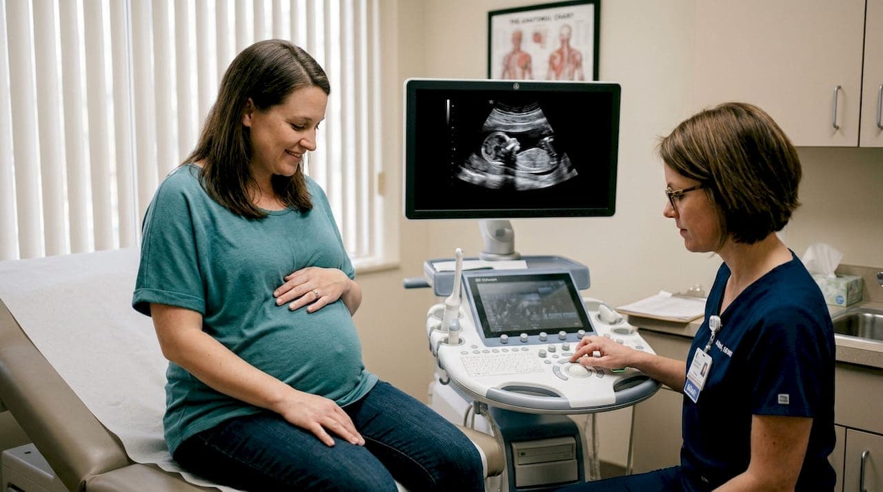
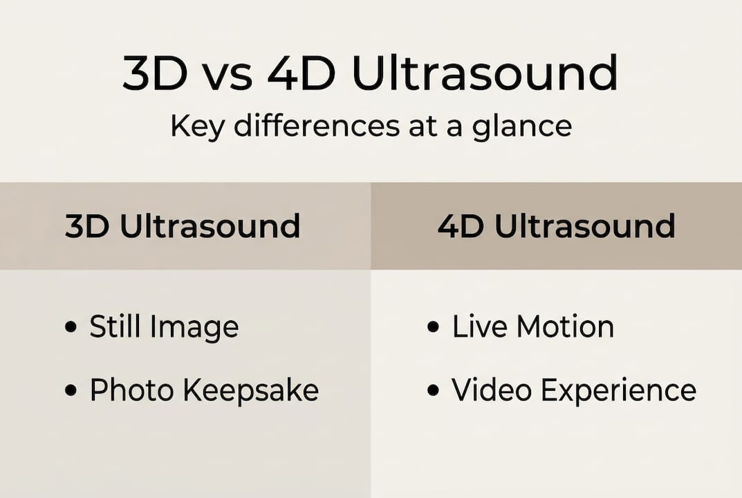
Before you can weigh your options, it helps to understand what you're actually comparing. Traditional 2D ultrasounds produce flat, grayscale cross-sections of your baby. They're the standard in most prenatal checkups. 3D ultrasounds go further by capturing multiple 2D images from different angles and combining them into a single still, three-dimensional image. You see shape, depth, and facial features frozen in one frame. 4D ultrasounds add the dimension of time, meaning you see that same three-dimensional image moving in real time, like a live video of your baby yawning, stretching, or opening their eyes.
The equipment needed for each type is more demanding than a standard machine. 3D/4D systems require advanced multi-dimensional transducers and high-processing hardware for volume acquisition and rendering, and 4D requires even higher frame rates to produce smooth motion. Not every clinic has this level of equipment, which matters when you're choosing where to go.
Here's a quick breakdown of how they compare at a foundational level:
| Feature | 3D Ultrasound | 4D Ultrasound |
|---|---|---|
| Image type | Still, three-dimensional | Live-motion video |
| Emotional impact | Single detailed snapshot | Real-time baby movement |
| Equipment demand | High | Very high |
| Best use case | Structural detail | Movement and bonding |
| Typical session length | 20-30 minutes | 30-45 minutes |
One thing both have in common is a medical framework. ACOG and AIUM recommend ultrasound only for medical benefit and discourage non-diagnostic or elective use based on a precautionary principle, even though risks are considered low. That doesn't mean elective 3D/4D scans are dangerous, but it does mean you should choose a certified provider rather than a pop-up keepsake shop.
Who benefits most from each? Parents who want a single stunning image to frame often prefer 3D. Parents who want to watch their baby move and capture that on video typically lean toward 4D. You can also explore the available ultrasound services to understand what's offered at specialized prenatal imaging centers before you decide.

Pro Tip: Drink at least 64 ounces of water daily in the week before your scan. Well-hydrated mothers tend to have more amniotic fluid, which dramatically improves image clarity for both 3D and 4D sessions.
Step-by-step: How to compare 3D and 4D ultrasound experiences
Now that you have the foundation, here's how to actually evaluate and compare your options before booking.
1. Assess image and video quality standards. Ask any provider you're considering to show you sample images or videos from their equipment. 3D still images should show clear facial features, visible skin texture, and defined contours. 4D footage should be smooth, not choppy or pixelated. If a clinic can't show you examples, that's a red flag.
2. Understand the diagnostic value of each type. Both have legitimate clinical uses beyond bonding. When it comes to complex structural assessments, 3D ultrasound showed 100% sensitivity, 72.7% specificity, and 90% accuracy in placenta accreta diagnosis, outperforming MRI in that specific application. For cardiac screening, a 4D meta-analysis found pooled sensitivity of 91%, specificity of 98%, and AUC of 0.98 for congenital heart defects (CHDs), with optimal results around 20 weeks. These numbers matter if you're considering a scan for medical monitoring.
3. Discuss safety and realistic expectations with your provider. Both 3D and 4D use the same ultrasound energy as 2D. The session just lasts longer. Make sure your provider explains what's possible given your gestational age and baby's position.
4. Choose timing carefully. Between 26 and 32 weeks is generally considered the sweet spot for bonding-focused scans. Earlier gives you a better structural view but less baby fat. Later gives more recognizable facial features.
Here's a side-by-side look at how they stack up across key decision factors:
| Decision factor | 3D | 4D |
|---|---|---|
| Best bonding format | Photo keepsake | Live video experience |
| Ideal gestational window | 24-28 weeks | 26-32 weeks |
| Diagnostic strength | Structural anomalies | Cardiac and movement |
| Rescheduling flexibility | Moderate | Often recommended |
You can also compare imaging technologies side by side through detailed breakdowns to help guide your final call.
Pro Tip: If your main goal is a beautiful keepsake, 4D tends to be more memorable because you capture movement. But if you want the clearest single image for a nursery print, a high-resolution 3D scan often wins.
Factors that affect image quality and outcome
Even the best machine and most experienced technician can't guarantee perfect images every time. Several real-world variables directly impact what you'll see on screen.
Physical factors you can't fully control:
- Fetal position: If your baby is facing your spine or has their hands covering their face, the image is limited. Providers often ask you to walk around or eat something sweet to encourage movement.
- Amniotic fluid levels: More fluid around the baby's face creates a clearer "window" for the scan. Low fluid can make images murky or incomplete.
- Maternal BMI: Higher body mass index can reduce ultrasound wave penetration, affecting depth and clarity regardless of machine quality.
- Placenta location: An anterior placenta (one that sits along the front of the uterus) can partially block the signal path.
Image quality can be limited by fetal position, amniotic fluid levels, and maternal BMI, which may lead to disappointment in elective settings if expectations aren't properly set beforehand.
Beyond physical factors, the expertise of your sonographer matters enormously. Operator skill is critical for complex assessments, especially when evaluating cardiac outflow tracts or other detailed structures where 3D and 4D outperform 2D. A technically advanced machine operated by someone without deep experience will produce inferior results compared to a slightly older machine in expert hands.
"The most common source of parent disappointment after a 3D/4D session isn't the technology, it's unmanaged expectations. The baby moved. The position was wrong. The fluid was low. Preparation and timing are everything."
To learn more about factors affecting ultrasound quality and how certified providers manage them, it helps to read up before your appointment.
Pro Tip: Schedule your elective 3D or 4D scan in the morning when you're well-rested and well-hydrated. Avoid carbonated drinks or large meals right before, as these can increase fetal activity unpredictably and reduce session control.
Common pitfalls and mistakes to avoid when choosing your scan
Knowing the technology is one thing. Avoiding the traps that trip up most parents is another.
1. Choosing a keepsake-only studio without medical oversight. These facilities often operate outside accreditation standards. The FDA and ACOG specifically caution against elective keepsake scans that expose the fetus to ultrasound without clinical justification. Always verify that the center employs registered diagnostic medical sonographers (RDMS).
2. Booking too early. Before 24 weeks, the baby hasn't developed enough subcutaneous fat to show defined facial features. Parents who book at 16 or 18 weeks often end up with skeletal-looking images and feel let down.
3. Booking too late. After 34 weeks, the baby is often too large and cramped to reposition. Head engagement and limited fluid make image quality unpredictable.
4. Confusing bonding with diagnosis. A 3D or 4D scan at a specialized center is not a substitute for your OB's anatomy scan. These are different tools for different purposes. One is about emotional connection, the other is clinical screening.
5. Not asking about rescheduling policies. Baby position and fluid levels are unpredictable. The best centers offer a free or low-cost return visit if your session doesn't produce quality results. Ask before you book.
"Parents who approach elective 3D/4D scans with realistic expectations and the right timing consistently report the experience as emotionally powerful. Those who don't often feel cheated, even when everything technically went fine."
Learning about what 3D and 4D offer at a certified center helps you set the right expectations from the start, which is often the difference between a magical experience and a frustrating one.
Pro Tip: Call ahead and ask specifically whether the center has a rebook or satisfaction policy. Reputable centers stand behind their sessions and will work with you if conditions weren't ideal.
The real value of 3D and 4D ultrasounds: Beyond the basics
Here's something most comparison articles won't tell you: the debate between 3D and 4D misses the bigger picture. After working with thousands of families over more than 15 years, what we've observed is that the technology is only half the story.
The other half is emotional readiness. Parents who arrive knowing what to expect, who've hydrated properly, who've scheduled at the right gestational window, and who've chosen a certified provider almost always leave with an experience that moves them deeply. Parents who arrive hoping for a perfect Instagram-worthy video regardless of baby's position often leave frustrated, even when the technology performed exactly as it should.
4D's motion can be breathtaking. Watching your baby yawn or wave in real time is unlike anything else in pregnancy. But a single well-timed 3D image, perfectly lit with HD Live technology, can stop a room. Neither is objectively better. The right choice depends on what kind of moment you want to create.
We've written about this extensively in insightful blog articles because we believe informed parents have better experiences. The most advanced scan in the world means nothing without the preparation and expertise behind it.
See your baby in a whole new way with advanced ultrasound
You now have a clear picture of what separates 3D from 4D, what affects your results, and how to avoid the most common mistakes. The next step is seeing the difference for yourself.
At BBview3D, our certified sonographers use HD Live technology across multiple U.S. locations to give your family an experience you'll treasure for life. Whether you're drawn to the depth of a 3D still or the moving magic of a 4D session, we offer packages built around your stage of pregnancy and your goals. You can explore all ultrasound services or see 3D/4D scan results from real families before you decide. First-time visitors also enjoy a limited-time discount on their initial appointment. Book with confidence, knowing you're in expert hands.
Frequently asked questions
Is a 4D ultrasound safer or more accurate than a 3D ultrasound?
Both are considered equally safe with low risk when performed by a qualified provider. ACOG recommends ultrasound only for medical benefit, and diagnostic accuracy between the two depends on the specific condition being evaluated and the timing of the scan.
When is the best time during pregnancy to schedule a 3D or 4D ultrasound?
For structural and cardiac screening, optimal results appear around 20 weeks, while bonding-focused sessions are generally best between 26 and 32 weeks when facial features are more developed.
What should I do if the images from my ultrasound are unclear?
Ask your provider about rescheduling. Image clarity depends on fetal position, amniotic fluid levels, and maternal BMI, and a second session with better preparation often produces significantly improved results.
Do insurance plans cover 3D or 4D ultrasounds?
Insurance typically covers ultrasounds only when there is a documented medical indication, meaning elective or keepsake sessions are generally paid out of pocket by the family.