Most expectant parents assume that every ultrasound appointment looks the same: a grainy, gray screen with a flickering shape that only a trained eye can read. That assumption undersells what modern technology can actually do for your family. Today's 3D and 4D prenatal imaging goes far beyond a quick medical check. It lets you see your baby's face, watch her yawn, and catch a tiny hand waving. This guide walks you through every major ultrasound feature, explains what each technology actually delivers, and helps you decide which experience is right for your family and your stage of pregnancy.
Table of Contents
- What makes a baby ultrasound? Types and technology explained
- Comparing 2D, 3D, and 4D ultrasounds: What you actually see
- Medical clarity vs. keepsake moments: Which uses are recommended?
- How 3D and 4D ultrasounds strengthen your emotional connection
- The hidden truth: Why informed parents get the best value from modern ultrasounds
- Take the next step: Experience advanced ultrasounds with BabyView3D
- Frequently asked questions
Key Takeaways
| Point | Details |
|---|---|
| Ultrasound types matter | 2D, 3D, and 4D ultrasounds each offer unique features and experiences for parents. |
| Safety is established | When used professionally, all modes are safe for both mom and baby. |
| Know when to choose | 3D/4D scans are best for added clarity, emotional connection, or when a doctor suggests. |
| Informed choices help | Being aware of medical guidance leads to the best balance of joy and reassurance. |
What makes a baby ultrasound? Types and technology explained
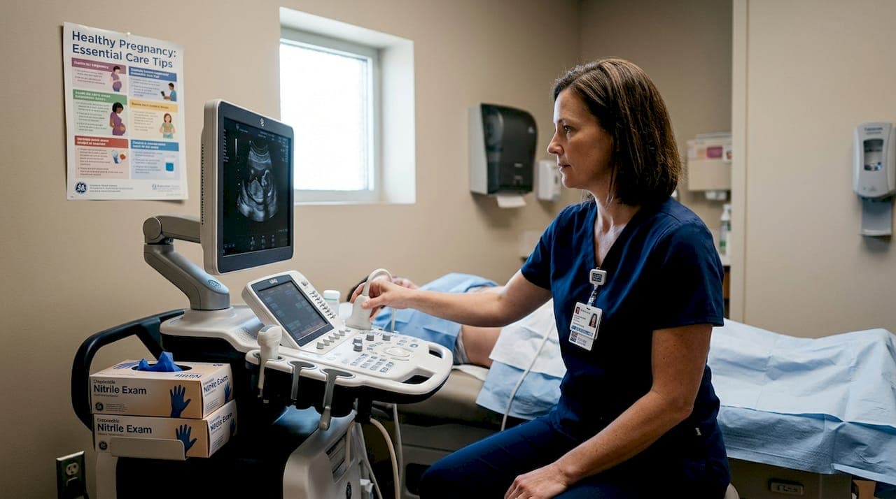
Ultrasound works by sending high-frequency sound waves into the body. Those waves bounce off tissue and fluid, return to the probe, and a computer translates the echo data into an image on screen. No radiation is involved. The process is the same whether your provider is using equipment from 1995 or a cutting-edge HD Live system.
What changes between 2D, 3D, and 4D is what the computer does with those echoes.
- 2D ultrasound captures flat, cross-sectional slices in real time. It is the gold standard for measuring your baby's growth and checking organ development.
- 3D ultrasound stacks hundreds of those slices and renders them into a static, three-dimensional surface image. You can see facial features, the curve of a nose, and the shape of tiny fingers.
- 4D ultrasound adds the element of time to a 3D render, producing live video so you can watch your baby move, stretch, and react in real time.
- 8K/HD Live imaging, offered at studios like BabyView3D, adds even richer color and texture mapping over the 4D render, making images look almost photographic.
A key safety point: 3D/4D ultrasounds use the same safe, non-ionizing sound wave technology as 2D, with no known risks when used appropriately, according to OB/GYN imaging safety guidelines. That means you can enjoy the emotional experience of a keepsake session without trading away peace of mind.
Recent ultrasound advancements have made 3D and 4D imaging available well outside the clinical setting, bringing professional-grade clarity into comfortable studio environments. You can explore the full range of ultrasound service types to understand what each modality offers.
| Type | Image style | Real-time? | Best use |
|---|---|---|---|
| 2D | Flat, grayscale | Yes | Medical checks, growth |
| 3D | Surface rendered, still | No | Facial detail, keepsakes |
| 4D | Surface rendered, live video | Yes | Movement, bonding |
| 8K/HD Live | Photorealistic color | Yes | Premium keepsake sessions |
Stat to know: Most elective 3D/4D studios recommend scheduling between weeks 26 and 32 for the clearest facial detail, though sessions are offered from 20 weeks onward.
Comparing 2D, 3D, and 4D ultrasounds: What you actually see
Now that we've covered how each ultrasound works, let's compare what you'll actually see and feel with each technology.
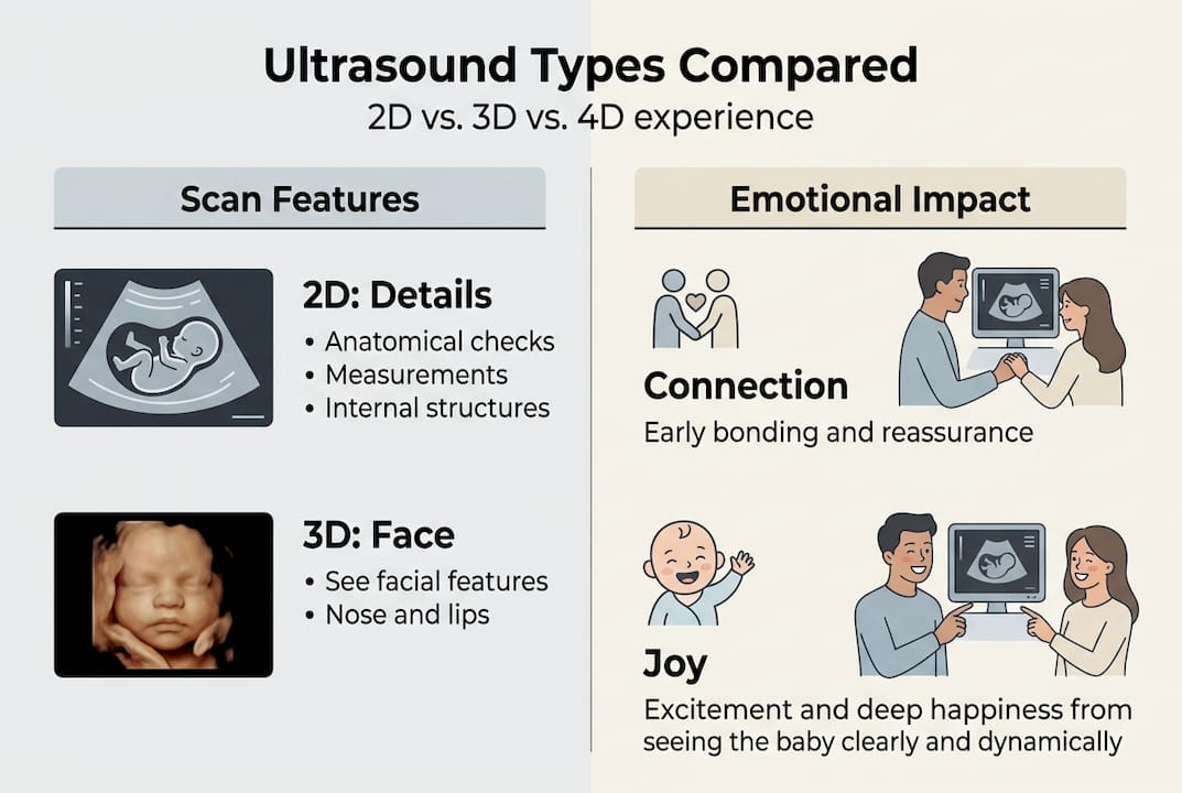
A 2D scan gives your provider exactly what they need medically: clear measurements, fluid levels, heartbeat confirmation, and organ placement. For parents, though, the image often looks abstract. You might make out a profile, but reading the screen typically requires guidance from your sonographer.
A 3D image changes everything emotionally. Suddenly, you're looking at a face. Cheeks, a button nose, lips slightly parted. Parents frequently describe the moment they see a 3D image as the point when their pregnancy became real. That emotional shift matters because it can strengthen attachment and help partners who have felt disconnected from the pregnancy start to bond in a tangible way.
4D takes it further. You're not looking at a still photo anymore. You're watching your baby in motion: a hand moving toward a mouth, a full-body stretch, eyelids fluttering. One parent described it as "watching a live feed of someone you already love." That is the kind of emotional memory that parents carry for years.
For diagnostic precision, 4D ultrasound shows pooled sensitivity of 91% and specificity of 98% in diagnosing fetal congenital heart disease, outperforming some 2D protocols. You can browse a full ultrasound image gallery to see examples before booking your session.
"Seeing our daughter's face in 4D before she was born made her feel completely real to us. Nothing could have prepared us for that moment."
Pro Tip: Drink 64 oz of water daily in the two days before your 3D/4D session. Proper amniotic fluid levels give the sound waves more room to render a clear, detailed image. A well-hydrated session produces noticeably sharper results.
| Modality | What parents see | Emotional impact | Optimal weeks |
|---|---|---|---|
| 2D | Flat shapes, organ outlines | Moderate | 8 to 40 |
| 3D | Facial features, still | High | 26 to 32 |
| 4D | Live movement, expressions | Very high | 26 to 32 |
| 8K HD Live | Photorealistic color video | Exceptional | 26 to 32 |
Medical clarity vs. keepsake moments: Which uses are recommended?
Now that you know what each ultrasound looks like, it's important to understand when each is used medically and when it's just for keepsake.
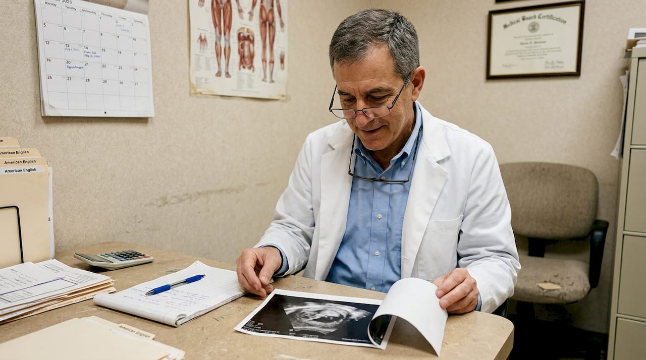
Your OB-GYN or midwife will use 2D ultrasound for every standard prenatal appointment. It measures the nuchal fold, checks placenta placement, monitors amniotic fluid, and confirms your due date. These are the scans that keep you and your baby safe throughout pregnancy.
3D and 4D are not part of routine prenatal care, and they were not designed to replace standard screening. 3D/4D is not routinely recommended by ACOG for standard prenatal care; 2D is sufficient for anatomy and growth checks, with 3D/4D serving as an adjunct for clarifying concerns like cleft lip or skeletal issues. You can review detailed ultrasound medical information to understand where each type fits in a prenatal care plan.
Here is when each type is typically used:
- 2D: Every prenatal visit, anatomy scan at 18 to 22 weeks, growth scans in the third trimester
- 3D: Suspected cleft lip or palate, further evaluation of limb or spine concerns, keepsake sessions
- 4D: Fetal cardiac evaluation, suspected movement disorders, emotional bonding sessions
- 8K/HD Live: Elective keepsake studio sessions for premium image and video quality
Pro Tip: If your provider recommends a 3D/4D scan for a medical reason, ask your OB whether the studio results will be reviewed by a certified sonographer and shared with your care team. A good studio will always support that communication.
For parents who want to book an elective keepsake session, the key is doing so in addition to your scheduled medical scans, not instead of them. The two experiences serve completely different purposes, and both have real value when used correctly.
Stat worth noting: Elective 3D/4D ultrasound studios in the U.S. operate separately from clinical settings and are not regulated under the same medical guidelines, which is why choosing a studio with certified sonographers matters enormously.
How 3D and 4D ultrasounds strengthen your emotional connection
Beyond medical uses, many parents seek ultrasounds to feel closer to their baby. Let's explore how modern imaging achieves this.
There is something powerful about seeing a face before you have ever held a person. Research in prenatal psychology shows that visualization plays a significant role in early attachment formation. When parents see a realistic image of their baby's features, the brain begins building a relational identity for that child. The pregnancy stops being abstract and becomes personal.
3D/4D imaging complements 2D by providing spatial context and reducing operator dependence, though it increases data complexity. This means you are getting a richer picture, but always works best alongside standard care rather than replacing it.
Here are steps to maximize the emotional impact of your 3D/4D session:
- Schedule at the right time. Weeks 26 to 32 offer the best balance of baby size and amniotic fluid space for clear facial imaging.
- Bring your support person. Sharing the experience live increases bonding for partners, grandparents, and siblings.
- Ask for a video keepsake. Watching the recording later, especially during sleepless nights after birth, reconnects you to that early wonder.
- Choose a studio with certified sonographers. Expertise matters for image quality and for answering your questions in the moment.
- Save everything. Digital files last. Printed ultrasound images fade over time, so store your 3D/4D video in the cloud.
Read real parent ultrasound stories to understand how other families have described their experience. The emotional clarity that comes from seeing your baby move in real time is something that many parents describe as one of the most memorable moments of their entire pregnancy journey.
The hidden truth: Why informed parents get the best value from modern ultrasounds
With this context, it's important to see the bigger picture. Here's a candid perspective on how parents can truly benefit from ultrasound technology.
The loudest marketing around 3D/4D ultrasounds focuses entirely on the emotional wow factor. And yes, it is wonderful. But we've seen, over 15 years of working with families, that the parents who walk away most satisfied are not the ones chasing the most dramatic experience. They are the ones who came in informed.
Informed parents ask the right questions: Is this studio staffed by certified sonographers? Does the equipment actually support HD Live rendering, or is it a marketing label? Will my session images be clear enough to share with my OB if needed?
The parents who overbuy without asking these questions sometimes end up disappointed. They book extra sessions thinking more scans equal better bonding, when in reality timing and preparation matter far more than volume. Check out expert ultrasound advice before you decide on a package. The goal is one great session, not four mediocre ones.
Combine emotional intention with informed decision-making, and you will get an experience worth remembering for the rest of your life.
Take the next step: Experience advanced ultrasounds with BabyView3D
For parents ready to bridge the gap between clarity, connection, and lasting memories, here's how you can get started.
At BabyView3D, we combine more than 15 years of experience with the most advanced HD Live imaging technology available. Our certified sonographers guide you through every moment of your session, making sure the images you leave with are as clear and meaningful as possible.
Whether you're looking to explore ultrasound packages that fit your timeline and budget, or you simply want to see real ultrasound images before committing, we make it easy to take that next step. First-time appointment offers and flexible session options are available when you visit BabyView3D and book online today.
Frequently asked questions
Are 3D and 4D ultrasounds safe for my baby?
Yes, 3D and 4D ultrasounds use the same safe, non-ionizing sound waves as 2D scans when performed appropriately. Safety is well established for both clinical and elective settings.
At what stage of pregnancy are 3D/4D ultrasounds most effective?
3D and 4D imaging is most effective between 20 and 32 weeks when your baby's features and movements are best captured. Optimal imaging occurs specifically in the 26 to 32 week window for facial detail.
Is a 3D or 4D ultrasound necessary for my routine prenatal care?
No, standard 2D ultrasound is sufficient for routine prenatal checks. 3D/4D is not routinely recommended by ACOG and is best used for additional clarity or keepsake purposes alongside your medical care.
Can a 3D or 4D ultrasound detect more conditions than 2D?
Yes, for specific conditions like heart defects, 4D ultrasound shows 91% sensitivity and 98% specificity, offering additional detail beyond standard 2D protocols for targeted clinical concerns.