
Not all ultrasounds are the same experience. While traditional 2D scans show flat, gray cross-sections that take trained eyes to interpret, 3D ultrasound produces vivid, surface-rendered images that let you actually see your baby's face, hands, and features months before birth. For many parents, that first real look at a tiny nose or curled fingers is one of the most emotionally powerful moments of pregnancy. This guide explains how 3D ultrasound works, what it does medically, what its real limitations are, and how to make smart decisions about your prenatal imaging choices.
Table of Contents
- What is 3D ultrasound? Defining the basics
- Clinical advantages: Medical benefits and accuracy
- Limitations and image quality: What parents should know
- Safety, emotional value, and choosing the right scan
- A new era: What 3D ultrasound really means for families
- See your baby in 3D: Next steps with BabyView3D
- Frequently asked questions
Key Takeaways
| Point | Details |
|---|---|
| Clear 3D imaging | 3D ultrasound delivers detailed, lifelike visuals that help parents connect with their baby before birth. |
| Medical advantages | Evidence shows 3D ultrasound offers high accuracy for diagnosing certain conditions compared to 2D. |
| Safety assurance | 3D ultrasound uses safe, non-ionizing sound waves, but should be performed by qualified professionals. |
| Know the limits | Image quality depends on timing, skill, and pregnancy stage—so not all scans provide picture-perfect results. |
| Choosing wisely | Elective 3D scans offer emotional value but must always prioritize medical guidance and provider qualification. |
What is 3D ultrasound? Defining the basics
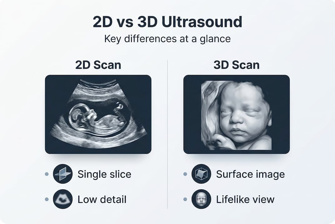
Standard 2D ultrasound captures single, flat slices of tissue using sound waves. You see a cross-section, like slicing a loaf of bread and looking at one piece. A 3D ultrasound collects hundreds or thousands of those flat slices rapidly, then uses specialized software to reconstruct them into a three-dimensional surface image. The result looks much more like a photograph than a diagram.
This technology has been advancing steadily, and understanding how the images are gathered helps you appreciate why quality varies so much between providers. There are three main acquisition methods used in modern 3D imaging:
- Freehand scanning: A standard probe is moved by hand while optical, electromagnetic sensors, or cameras track its position precisely. The software then assembles slices based on tracked movement data.
- Mechanical probes: These contain internal motors that rotate or wobble the transducer automatically, sweeping through tissue in a controlled arc without requiring the operator to move the probe manually.
- Matrix array probes: These use hundreds of tiny transducer elements arranged in a two-dimensional grid. By electronically steering sound beams in multiple directions simultaneously, they capture volumetric data almost instantly.
The method used makes a real difference. Matrix array probes tend to produce the most consistent, detailed volumes because the data capture is fast and doesn't depend on smooth hand movement. Freehand systems offer flexibility but are more operator-dependent.
Here's a simple comparison to put the technologies side by side:
| Feature | 2D ultrasound | 3D ultrasound |
|---|---|---|
| Image output | Flat, single-plane slice | Surface-rendered 3D volume |
| Baby's features visible? | Rarely, requires training | Yes, clearly visible |
| Data capture method | Continuous single beam | Multiple slices reconstructed |
| Probe types | Standard transducer | Mechanical, freehand, matrix array |
| Clinical use | Routine screening | Specialized diagnostics and elective |
| Emotional impact for parents | Limited | Very high |
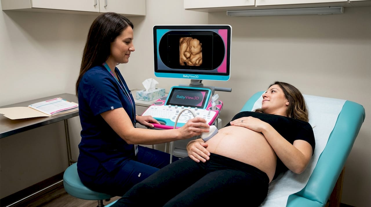
Exploring 3D ultrasound technology further reveals why the viewing experience is so different. With 2D, parents typically need a technician to point out "there's the head" or "that's the spine." With 3D, you can recognize your baby's profile on your own. That shift from medical data to visible person is what makes this technology so meaningful, both clinically and emotionally.
If you want to see what modern imaging actually looks like in practice, reviewing ultrasound service offerings from experienced providers gives a real sense of what current technology can achieve.
Clinical advantages: Medical benefits and accuracy
Now that you know how it works, let's look at why it's medically meaningful, especially when diagnostic accuracy matters most.
3D ultrasound is not just a keepsake tool. It has genuine, evidence-based medical applications that in some situations outperform both traditional 2D ultrasound and even more expensive imaging modalities like MRI.
One of the strongest examples is placenta accreta spectrum, a serious condition where the placenta attaches too deeply into the uterine wall. Getting this diagnosis right before delivery can be life-saving. Research comparing diagnostic methods found that 3D ultrasound reached 100% sensitivity, 72.7% specificity, and 90% overall accuracy for diagnosing placenta accreta spectrum in a study of 60 patients. In other words, it correctly identified every true case, which is a remarkable result for any imaging technology.
Here's how those numbers stack up in context:
| Diagnostic measure | 3D ultrasound result |
|---|---|
| Sensitivity | 100% |
| Specificity | 72.7% |
| Overall accuracy | 90% |
| Study population | 60 patients |
Beyond serious complications, 3D ultrasound is also proving valuable in the routine precision measurements taken early in pregnancy. Measuring the crown-rump length (CRL) in the first trimester helps calculate due dates and assess fetal growth. Traditionally this takes a trained sonographer about 35 seconds per measurement. An AI-enhanced 3D method achieves 91.4% standard plane accuracy in just 5 seconds, with a mean absolute error of only 1.58mm compared to a 2D expert baseline, across a 245-patient external validation group. That speed and accuracy together represent a significant step forward.
Here's why these clinical advantages matter for expectant parents specifically:
- Early detection of structural concerns: Surface rendering helps identify facial differences, limb anomalies, and neural tube issues more clearly than 2D alone.
- Improved anatomy surveys: Organ positioning, spine integrity, and palate structures are more clearly visualized in three dimensions.
- Better communication with your care team: When your doctor can show you a 3D image of a finding, it's far easier to understand what's being discussed and to make informed decisions.
- Reduced need for repeat scans: Because 3D captures volumetric data, stored images can be reviewed and measured after the scan is complete, without requiring you to come back.
Reading more about medical ultrasound guidelines can help you understand which clinical scenarios are most likely to benefit from 3D imaging in your specific pregnancy.
Limitations and image quality: What parents should know
Even with clinical strengths, it's vital to understand the practical side, and why optimal results aren't guaranteed for every scan.
The most important thing to understand is this: 3D image quality is entirely dependent on 2D image quality. The software reconstructs a three-dimensional volume from flat slices, so if the underlying 2D images are poor, the 3D result will be too. There is no software workaround for bad raw data.
Several factors directly affect what you'll see:
- Baby's position: If the baby's face is pressed against the placenta or toward your spine, clear facial images are simply not possible.
- Amniotic fluid levels: The fluid acts as a natural window for sound waves. Low fluid reduces clarity significantly.
- Gestational age: Very early in pregnancy, structures are too small and undifferentiated for meaningful 3D rendering.
- Maternal body composition: Higher levels of abdominal tissue can scatter and weaken the sound waves before they reach the baby.
- Probe type and provider skill: As noted earlier, the acquisition method and the operator's technique both directly shape the final image quality.
The 3D ultrasound Radiopaedia resource notes that endovaginal scanning produces better results for gynecological applications, while transvaginal limitations mean early pregnancy 3D scans (before 14 weeks) rarely yield impressive images. This is worth knowing if you're planning an early elective session.
It is also important to understand that 3D ultrasound is not routine care for healthy pregnancies. Standard prenatal screening still relies on 2D imaging, and your OB or midwife's clinical scans are the primary medical record of your baby's development. 3D is a complement, not a replacement.
"The best 3D session is the one you plan thoughtfully, at the right gestational age, with a skilled provider, and with realistic expectations about what you'll see."
Pro Tip: Call ahead and ask your chosen provider what happens if the baby isn't cooperating during your session. Reputable studios will offer to reschedule or extend your time, not just send you home with blurry screenshots.
Working with qualified providers who are honest about what's achievable in your specific situation makes all the difference between a disappointing experience and a deeply moving one.
Safety, emotional value, and choosing the right scan
With limitations discussed, let's close the loop by answering parents' biggest concerns, including safety, emotional impact, and practical next steps.
Is 3D ultrasound safe?
This is the first question almost every parent asks, and the answer is reassuring. 3D ultrasound uses the same non-ionizing sound waves as standard 2D ultrasound. There is no radiation involved. When performed by trained professionals in medically indicated scenarios, it is considered safe. That said, professional health organizations caution against prolonged or purely commercial elective use that has no diagnostic purpose, simply because extended unnecessary exposure, even with safe technology, is never the recommended standard.
The key takeaway: safety is about context and provider quality, not the technology itself.
The emotional value is real and significant
Parents who see their baby's face clearly in 3D often describe a profound shift in how real the pregnancy feels. Studies in perinatal psychology suggest that early parent-to-baby bonding has lasting positive effects on maternal behaviors and family attachment. Seeing a yawn, a stretch, or a tiny hand pressed against a cheek creates a connection that no flat, gray image can replicate. This isn't a marketing claim. It's a consistent finding from families across every demographic.
Choosing the right time and provider
Here's a practical guide to getting the most from your 3D experience:
- Schedule between 24 and 32 weeks. This window offers the best combination of developed baby features and sufficient amniotic fluid. After 32 weeks, the baby becomes more cramped and fluid decreases, which can limit image clarity significantly.
- Ask about equipment. Matrix array probes and HD Live rendering systems produce noticeably better images than older mechanical probe systems.
- Check credentials. Your sonographer should be registered or certified. At minimum, ask how long they've been performing 3D sessions specifically, not just 2D clinical scans.
- Drink water beforehand. Being well-hydrated improves amniotic fluid levels and generally improves acoustic windows.
- Choose providers who offer rescheduling. If the baby isn't positioned well, a trustworthy studio accommodates that reality.
Pro Tip: Avoid providers who guarantee specific images or make promises about facial clarity. Reputable sonographers are upfront about the variables involved, because that honesty is exactly what makes them trustworthy.
For guidance on scheduling and what to expect from your session, ultrasound guidance from experienced professionals can help you prepare with confidence.
A new era: What 3D ultrasound really means for families
Here's something the standard clinical literature doesn't say directly but that fifteen-plus years of family imaging makes clear: 3D ultrasound is not primarily a medical tool that happens to be emotional. It's a fundamental shift in what pregnancy looks like to the people living it.
For most of human history, parents met their babies at birth. Then 2D ultrasound arrived and gave parents a blurry signal that something was there. Now 3D imaging lets you recognize your baby, weeks before delivery. That is a genuinely new experience in the history of human parenthood, and its implications for bonding, preparation, and family connection are still being understood.
What concerns us at BabyView3D is not the technology itself but the gap between what's possible and what families actually receive. Too many parents book sessions based on price alone and end up with poor images from undertrained operators using outdated equipment. That experience doesn't just disappoint. It can actually reduce trust in prenatal imaging altogether.
The other uncomfortable truth is that not every 3D session will produce a beautiful image, and any provider who implies otherwise isn't being straight with you. The variables are real. But when the timing is right, the provider is skilled, and the technology is current, the experience of seeing your baby in three dimensions is something you simply can't replicate through any other means.
We encourage every family to read parent perspectives on 3D imaging, ask hard questions before booking, and choose providers whose reputation is built on honest, skilled work rather than flashy marketing. The technology deserves that level of care. So does your family.
See your baby in 3D: Next steps with BabyView3D
If this guide has answered your questions and you're ready to experience the difference that professional 3D imaging makes, BabyView3D is here to make it exceptional.
With over 15 years of experience, certified sonographers, and the latest HD Live imaging technology across multiple U.S. locations, BabyView3D specializes in creating the kind of experience you'll remember forever. Every session is designed around your family, your baby, and the moments that matter most. Explore our full range of BabyView3D services to find the package that fits your pregnancy stage and your vision. And before you book, take a look at our baby 3D ultrasound gallery to see exactly what current technology can capture. Your baby's face is waiting to be seen.
Frequently asked questions
Is 3D ultrasound safe for my baby?
Yes. 3D ultrasound uses the same non-ionizing sound waves as regular 2D ultrasound and is considered safe when performed by qualified professionals in medically indicated or elective settings managed by trained sonographers.
When is the best time in pregnancy for a 3D ultrasound?
The optimal window is 24 to 32 weeks, when baby features are well-developed, amniotic fluid is plentiful, and imaging conditions are most favorable for clear, detailed results.
Can 3D ultrasound detect medical conditions earlier than 2D?
In specific clinical scenarios, yes. 3D ultrasound demonstrated 100% sensitivity and 90% accuracy for diagnosing placenta accreta spectrum, outperforming standard 2D evaluation in that study population.
Are there situations where 3D ultrasound is not recommended?
3D ultrasound has limitations in early pregnancy and in cases with poor acoustic windows. It is not part of routine prenatal care for low-risk pregnancies and should be used where clinically appropriate or thoughtfully for elective bonding sessions.
How do I choose a qualified 3D ultrasound provider?
Look for certified sonographers with specific 3D experience, ask about the equipment used, and choose providers who are transparent about what results are realistic rather than those who over-promise image outcomes.