Most expectant parents walk into their first ultrasound expecting a grainy, black-and-white blur that vaguely resembles a baby. What they actually see today stops them in their tracks. Modern prenatal imaging has advanced so dramatically that families now watch their baby yawn, stretch, and even smile in real time, all before birth. Advanced imaging technology visually connects expectant parents with their developing baby in ways that were simply impossible a generation ago. This article breaks down how these tools work, what they mean for your pregnancy journey, and why the experience matters far beyond the technical specs.
Table of Contents
- How ultrasound technology works: From soundwaves to vivid images
- Innovations that shape your experience: AI and enhanced imaging
- What parents see and feel: The emotional power of advanced ultrasounds
- Safety, limits, and future trends in ultrasound technology
- Our perspective: Why parental memories matter more than megapixels
- Experience advanced ultrasounds and create special memories
- Frequently asked questions
Key Takeaways
| Point | Details |
|---|---|
| Advanced imaging | Modern ultrasounds now provide lifelike views, making it easier to connect with your baby before birth. |
| AI-powered accuracy | New technology improves measurement precision and helps predict delivery dates faster than ever. |
| Safety assured | Diagnostic ultrasounds are safe for parents and babies when performed responsibly. |
| Lasting memories | 3D/4D ultrasounds create emotional bonds and keepsakes for families to cherish. |
| Smart use of tech | Using ultrasound technology thoughtfully enhances the pregnancy experience without unnecessary scans. |
How ultrasound technology works: From soundwaves to vivid images
Now that you know ultrasounds reveal far more than you might imagine, let's look at the fascinating technology making this possible.
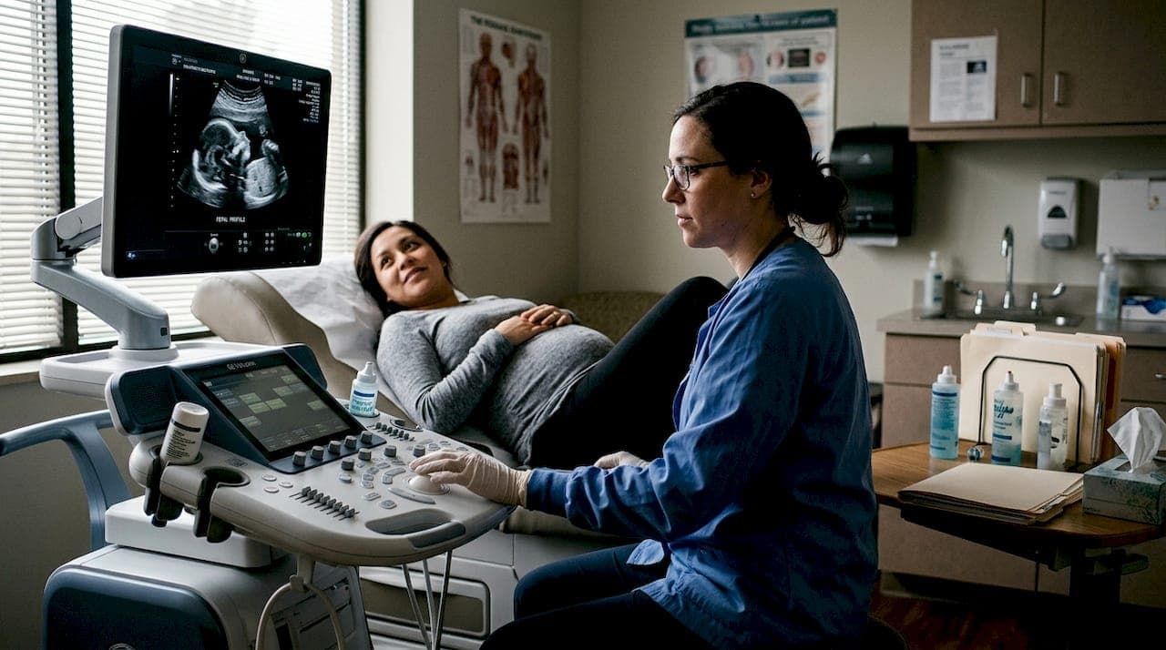
At its core, an ultrasound machine sends high-frequency sound waves into the body and listens for the echoes that bounce back. Those echoes are then translated into images on a screen. There is no radiation involved, which is one reason ultrasound has been the go-to imaging tool in pregnancy for decades.
Piezoelectric transducers convert electrical energy into high-frequency sound waves that reflect off fetal tissues to create images. The transducer, the handheld device pressed against your belly, both sends and receives these waves. The speed and strength of the returning echoes tell the machine how dense or soft each tissue is, building a picture layer by layer.
Here is a quick look at how the main scanning modes compare:
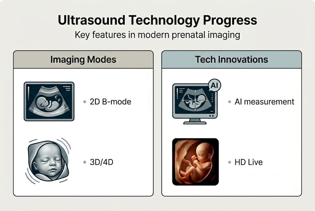
| Scanning mode | Frequency range | Probe type | What you see |
|---|---|---|---|
| 2D B-mode | 2 to 18 MHz | Linear or curved | Flat, grayscale cross-sections |
| 3D | 3 to 9 MHz | Motorized array | Still, three-dimensional surface images |
| 4D | 3 to 9 MHz | Motorized array | Live, moving 3D video |
The jump from 2D to 3D and 4D is not just cosmetic. Motorized array probes sweep through tissue at rapid angles, capturing hundreds of image slices per second. Software then stitches those slices together into a lifelike, three-dimensional view. You can see your baby's nose, cheeks, and fingers with striking clarity.
Key advantages of advanced scanning modes include:
- Real-time movement captured in 4D lets parents see facial expressions as they happen
- Surface rendering in 3D highlights skin texture and facial features with impressive detail
- Improved depth perception helps sonographers spot structural details more accurately
- Reduced scan time with motorized probes means less time lying still for you
For a deeper look at comparing imaging modes side by side, it helps to understand what each format is best suited for before your appointment. If you want to know more about how 3D/4D ultrasounds work in a clinical setting, that context will help you ask smarter questions.
Pro Tip: Before booking any ultrasound session, ask specifically which probe technology and rendering software the provider uses. Not all 3D/4D machines are created equal, and the difference in image quality can be significant.
Innovations that shape your experience: AI and enhanced imaging
Understanding the basics sets the foundation. Next, let's see what makes the latest ultrasounds so impressive and parent-friendly.
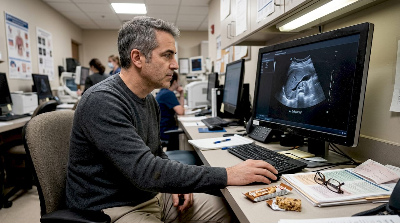
Artificial intelligence is no longer a futuristic concept in prenatal care. It is already inside the machines being used today. AI-powered ultrasound software can automatically identify anatomical landmarks, measure fetal head circumference, femur length, and abdominal size, and calculate gestational age in seconds. Tasks that once required several minutes of careful manual work now happen almost instantly.
The accuracy is genuinely impressive. AI automates fetal measurements, improves accuracy, and has received FDA clearance for delivery date estimation, with predictive models achieving an R² accuracy of 0.92. That means the system's due date predictions align very closely with actual delivery dates across large patient populations.
Image enhancement is the other major leap forward. Radiant mode and AI-assisted imaging help in challenging cases and reduce operator-dependent differences, meaning the quality of your images depends less on which sonographer happens to be in the room that day.
Here is how the generations of ultrasound technology compare:
| Feature | Traditional 2D | 3D/4D standard | AI-assisted ultrasound |
|---|---|---|---|
| Image type | Flat, grayscale | 3D surface rendering | Enhanced, auto-optimized |
| Measurements | Manual, time-consuming | Semi-automated | Fully automated |
| Due date accuracy | Moderate | Moderate | High (R² 0.92) |
| Operator dependency | High | Moderate | Low |
| Parent experience | Limited visual detail | Rich visual detail | Rich detail plus faster results |
Here is what a typical enhanced ultrasound session looks like step by step:
- Check-in and preparation: You arrive, drink water as directed, and the sonographer reviews your pregnancy history.
- Gel application: A warm gel is applied to your abdomen to help the probe make full contact with your skin.
- Initial 2D sweep: The sonographer takes standard measurements and checks fetal position.
- AI measurement phase: The software automatically identifies key anatomical points and logs measurements.
- 3D/4D rendering: The probe captures the surface view, and the image is rendered in real time on screen.
- Review and keepsakes: You watch the live session, ask questions, and receive your images or video.
For the latest on ultrasound advancements in prenatal care, staying informed helps you choose the right experience for your family.
What parents see and feel: The emotional power of advanced ultrasounds
Having looked at what happens during the scan, let's explore why these images matter so much on a personal level.
There is a moment during a 4D ultrasound when a baby opens its mouth or reaches up toward its face, and the room goes quiet. Parents often describe it as the moment pregnancy became real. That reaction is not accidental. It is the direct result of technology that now renders your baby's face with enough clarity to recognize family resemblances.
3D and 4D visualization supports emotional bonding and provides realistic, reassuring views of the baby that standard 2D scans simply cannot match. For parents who feel disconnected during pregnancy, especially in the early or middle trimesters, seeing a lifelike image can be genuinely grounding.
The emotional benefits parents report most often include:
- Stronger early bonding with the baby before birth, which research links to positive postpartum outcomes
- Reduced pregnancy anxiety when parents can visually confirm the baby is active and developing
- Shared experience with partners, grandparents, and siblings who can all watch the session together
- Lasting keepsakes in the form of printed photos, USB videos, and digital files families treasure for years
"When parents see their baby's face clearly for the first time, something shifts emotionally. That image becomes an anchor for the entire pregnancy and beyond." — Prenatal imaging specialist
Advanced 3D/4D ultrasound experiences are also increasingly used to help parents prepare emotionally for babies with known medical considerations, giving them a visual connection even in complex situations. It is worth noting that elective scans should always be performed by trained sonographers who monitor image quality and session length appropriately.
Safety, limits, and future trends in ultrasound technology
Now that you understand the emotional and experiential side, it is essential to consider safety, ongoing limitations, and how the future may change ultrasound experiences.
Ultrasound has one of the strongest safety records of any medical imaging tool. Ultrasound is safe at diagnostic levels when thermal and mechanical indices are monitored, but non-essential scans should be limited. Those two indices measure how much heat and pressure the sound waves generate in tissue. Reputable providers keep both well within established safe ranges.
That said, there are real limits and cautions worth knowing:
- Overuse is discouraged: Repeated elective sessions with no clinical purpose are not recommended by major medical organizations
- Position matters: A baby facing away from the probe or with limited amniotic fluid can result in poor image quality regardless of machine quality
- Not diagnostic: Elective 3D/4D sessions are not substitutes for clinical anatomy scans performed by a physician or certified diagnostic sonographer
- Operator skill still counts: Even with AI assistance, the person holding the probe shapes the session significantly
- Edge case limitations: Very high BMI or low fluid levels can reduce image clarity even with the best equipment
Looking ahead, AI promises personalized scan reports, faster session times, and even predictive health flags based on fetal measurements. But AI in ultrasound also has real limitations, including device variability across manufacturers and the risk of biased results in underrepresented patient populations.
For balanced ultrasound safety insights that go beyond the basics, it helps to read from providers who are transparent about both the benefits and the boundaries of the technology they use.
Pro Tip: Always ask your provider whether AI is being used during your scan, who interprets the final images, and whether a certified sonographer is present throughout the session. Transparency here is a sign of a trustworthy practice.
Our perspective: Why parental memories matter more than megapixels
After all the facts and features, let's step back and look at what really matters in your ultrasound journey.
We have spent over 15 years watching families walk out of ultrasound sessions holding a printed image like it is the most important thing in the world. And honestly, it is. Not because of the resolution or the AI-powered rendering behind it, but because of what it represents: the first time they truly saw their baby.
Here is the uncomfortable truth about ultrasound technology: most parents cannot tell the difference between a good 3D machine and a great one. What they remember is whether they felt welcomed, whether the sonographer took time to explain what they were seeing, and whether they left with something they wanted to show everyone they know.
Technology is a tool. A powerful one, yes. But chasing the highest megapixel count or the newest AI feature misses the point entirely. The families who treasure their ultrasound experience most are the ones who were fully present in the room, not the ones who had the fanciest equipment.
We encourage you to use technology as a bridge to connection, not a goal in itself. Explore memorable 3D/4D ultrasound packages that prioritize your experience as much as the image quality.
Experience advanced ultrasounds and create special memories
Ready to see your baby and keep that memory forever? Here is how you can experience every benefit discussed above.
At BabyView3D, we use state-of-the-art ultrasound technology including HD Live and 8K resolution imaging to give you the clearest, most detailed views of your baby available today. Our certified sonographers bring over 15 years of experience to every session, making sure you feel informed, comfortable, and genuinely moved by what you see.
Whether you are looking to explore ultrasound services for the first time or ready to shop keepsake packages that let you share this moment with everyone you love, we have options designed around your family. First-time visitors can take advantage of our limited introductory offer. Book your session today and see your baby like never before.
Frequently asked questions
Are 3D and 4D ultrasounds safe for my baby?
Yes, when performed as recommended, 3D and 4D ultrasounds are considered safe. Overuse is discouraged, but there is no confirmed harm at diagnostic levels when sessions are conducted by trained professionals.
What makes AI-powered ultrasounds different?
AI-powered ultrasounds automate measurements and speed up the session significantly. AI improves accuracy and can predict your due date with an R² accuracy of 0.92, which is notably higher than traditional manual methods.
How soon can I see realistic images of my baby?
Most families get the clearest 3D/4D images between 24 and 32 weeks of pregnancy. Advanced ultrasound technology makes lifelike images possible, though your baby's position and fluid levels also play a role.
Does insurance cover advanced ultrasounds for keepsakes?
Most insurance plans only cover ultrasounds that are medically necessary. Elective 3D/4D keepsake sessions are typically considered out-of-pocket expenses, so it is worth checking your specific plan before booking.