
Walking into an ultrasound appointment is one of the most exciting moments of pregnancy. But when the screen lights up with swirling gray shapes and your sonographer starts pointing at things you can barely make out, that excitement can quickly mix with confusion. You want to feel connected to your baby, not lost in a sea of pixels. The good news is that learning to read ultrasound images is not as complicated as it looks. This guide walks you through the types of scans, what to look for at each stage, how to decode your images step by step, and how to handle the moments when things seem unclear.
Table of Contents
- Understanding types of ultrasound and what each shows
- Essential components you'll see on an ultrasound
- Step-by-step: Interpreting your ultrasound images at home
- Troubleshooting unclear images and common pitfalls
- What happens next: Following up and capturing the memory
- A fresh perspective on what ultrasound interpretation really means
- Bring your ultrasound memories to life with BabyView3D
- Frequently asked questions
Key Takeaways
| Point | Details |
|---|---|
| Interpreting scans builds confidence | Learning the basics of ultrasound images empowers you to connect more deeply with your baby. |
| Most flagged findings are harmless | Over 80% of abnormalities noted in scans result in healthy outcomes after follow-up. |
| 3D and 4D open new perspectives | Advanced scans offer more detailed views and memorable moments for expectant families. |
| Ask for clarification | If you have questions or concerns about your scan, your provider welcomes follow-up discussions. |
Understanding types of ultrasound and what each shows
Before you can interpret an image, you need to know what kind of image you're looking at. Not all ultrasounds are the same, and each type gives you a different window into your baby's world.
2D ultrasound is the standard scan most parents see at their OB appointments. It produces flat, grayscale cross-sections of the body. Think of it like a slice through your baby, similar to how you'd cut through a loaf of bread to see the inside. It's the workhorse of prenatal care because it clearly shows internal structures like the heart, brain, and spine.
3D ultrasound takes hundreds of 2D slices and stitches them together to create a surface-rendered image. Instead of a cross-section, you see a three-dimensional shape, often revealing your baby's face, hands, and feet with striking clarity. Clinically, advanced ultrasound imaging using multiplanar views in 3D helps detect anomalies like cleft lip by showing the face from multiple angles simultaneously. Standardized ultrasound protocols ensure that each anatomy survey covers the head, heart, and abdomen in a consistent way, as outlined in OBUS Standards.
4D ultrasound adds the dimension of time to a 3D image, giving you live, moving footage of your baby. You might catch a yawn, a stretch, or even a smile. It's the closest thing to meeting your baby before birth.
Here's a quick comparison to keep things straight:
| Type | Image style | Best for | What you'll see |
|---|---|---|---|
| 2D | Flat grayscale | Medical assessment | Internal organs, measurements |
| 3D | Surface rendering | Facial features, anatomy | Face, hands, feet |
| 4D | Live 3D motion | Real-time movement | Baby moving, expressions |
When reviewing any scan, look for these key structures depending on your trimester:
- First trimester: gestational sac, yolk sac, fetal pole, heartbeat flicker
- Second trimester: brain, spine, heart chambers, face, limbs
- Third trimester: detailed facial features, fat deposits, practice breathing movements
Knowing which type of scan you have in hand is your first step toward confident interpretation.
Essential components you'll see on an ultrasound
Once you know your scan type, the next step is learning what to look for at each stage of pregnancy. The structures visible on ultrasound change dramatically as your baby grows.
In early pregnancy, your sonographer focuses on confirming a healthy start. The gestational sac appears around weeks 4 to 5, the yolk sac follows shortly after, and the fetal pole becomes visible around week 6. Cardiac activity is typically detectable between weeks 6 and 7, appearing as a tiny, rapid flicker on screen. Crown-rump length, or CRL, is the measurement from the top of your baby's head to the bottom of their spine, and it's the most accurate dating method in the first trimester up to 13 weeks and 6 days.

Here's a simple trimester timeline for what becomes visible:
| Week range | Structures visible |
|---|---|
| 4 to 6 weeks | Gestational sac, yolk sac, fetal pole |
| 6 to 7 weeks | Heartbeat, early fetal shape |
| 11 to 13 weeks | Head, limb buds, nuchal fold |
| 18 to 22 weeks | Brain, spine, heart, face, sex |
| 28 to 40 weeks | Fat, detailed features, breathing |
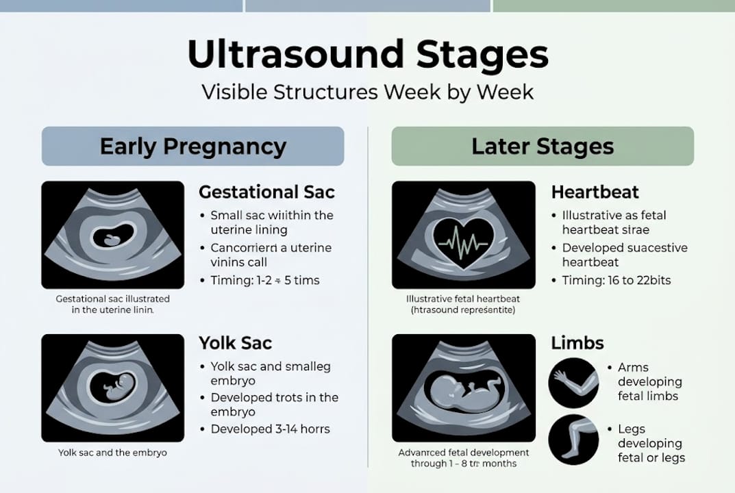
For the second trimester anatomy scan, sonographers follow a head-to-toe checklist. They measure the head circumference, look at brain structures, check the four chambers of the heart, examine the spine for closure, and count limb segments. By the third trimester, the focus shifts to growth, fluid levels, and position.
Here's a numbered approach to spotting structures on your own images:
- Find the largest, most defined shape. In early scans, that's usually the gestational sac.
- Look for a bright white oval inside it. That's the yolk sac.
- Spot the small elongated shape attached to the sac. That's your baby.
- Notice any rapid flickering movement. That's the heartbeat.
- In later scans, trace the bright white curved line. That's the spine.
Pro Tip: Don't stress if structures appear faint or incomplete in early images. An embryo at 6 weeks is only about 5 millimeters long, roughly the size of a pomegranate seed. Faint doesn't mean wrong.
Exploring ultrasound stages with a certified sonographer makes these details far easier to spot in real time.
Step-by-step: Interpreting your ultrasound images at home
With an understanding of ultrasound basics, here's how to decode your images step by step.
When you receive printed or digital images from your appointment, resist the urge to zoom in on one area and panic. Instead, use a systematic approach.
- Confirm the basics. Check the patient name, date, and gestational age printed on the image. This tells you exactly what stage you're looking at.
- Find the orientation markers. Most images show a label like "R" for right or an arrow indicating the top of the image. This helps you know which direction is which.
- Identify the largest structure. In early scans, it's the uterus or sac. In later scans, it's the baby's body.
- Look for bright white areas. Bone reflects sound waves strongly and appears bright white. This is how you spot the skull, spine, and limbs.
- Find dark areas. Fluid appears nearly black. The amniotic fluid surrounding your baby is a helpful reference point.
- Locate the heart. In 2D scans, look for a small, rhythmically moving area in the chest. In Doppler images, color overlays (usually red and blue) show blood flow direction.
For 3D and 4D images, look for the curved surface of your baby's face. The nose, lips, and eye sockets are often clearly visible. Hands appear as small, fan-shaped clusters of bright lines.
"Between 15 and 25 percent of ultrasounds are flagged as having something to follow up on, but 80 to 85 percent of those resolve as completely normal variations."
Pro Tip: Don't panic over isolated "abnormal" notations on your report. Radiologists are trained to flag anything that could possibly need attention, which means they intentionally over-report to avoid missing real issues. A flag is a prompt for conversation, not a diagnosis.
For more context on reading your results, ultrasound best practices can help you prepare smart questions for your provider.
Troubleshooting unclear images and common pitfalls
Sometimes, even with the best preparation, images are confusing or unclear. Here's how to handle that.
Unclear ultrasound images are far more common than most parents realize. Several factors can affect image quality:
- Fetal position: If your baby is facing away or curled tightly, key structures may be hidden.
- Amniotic fluid levels: Low fluid reduces the acoustic window, making images harder to read.
- Maternal body type: Higher body mass index can reduce sound wave penetration, softening the image.
- Gestational age: Very early scans simply don't have enough fetal tissue to produce crisp images.
- Artifact: Aliasing (where high-velocity blood flow appears as mixed colors in Doppler), shadowing from bone, and reverberation can all create misleading patterns.
According to indeterminate findings data, between 50 and 60 percent of scans contain some minor ambiguity. That's the majority of scans. It does not mean something is wrong.
"Indeterminate findings are common and rarely mean something harmful."
When you encounter an unclear image, here's what to do:
- Write down the specific area or notation that concerns you before your next appointment.
- Ask your provider whether a follow-up scan is clinically indicated or simply optional for reassurance.
- Request that your sonographer explain what they saw in plain language, not medical shorthand.
- Avoid searching individual findings online without full context. Isolated symptom searches almost always lead to worst-case scenarios.
Pro Tip: Jot down your questions the moment you look at your images at home. By the time your next appointment arrives, you'll have a focused list rather than a vague sense of worry.
For a broader look at how providers approach ambiguous results, common ultrasound challenges offers helpful context.
What happens next: Following up and capturing the memory
Once you've interpreted your ultrasound images, you may wonder what comes next, both medically and in making memories.
On the medical side, your provider will recommend next steps based on what the scan showed. A completely normal anatomy scan typically means no follow-up imaging until your third trimester growth scan. If something was unclear or flagged, your provider may recommend a targeted scan with a maternal-fetal medicine specialist, additional blood work, or simply a repeat scan in two to four weeks to recheck a specific structure.
Standardized anatomy protocols ensure that follow-up scans cover the same systematic checklist, so nothing gets missed between appointments.
On the memory side, your ultrasound images are among the first photos you'll ever have of your child. Here are some meaningful ways to preserve them:
- Digital albums: Organize images chronologically in a shared cloud folder so grandparents and family can follow along.
- Printed keepsakes: High-resolution 3D prints can be framed or included in a baby book.
- Announcement cards: Many parents use profile shots from 3D scans for pregnancy announcements.
- Video compilations: 4D session recordings can be edited into short clips to share at baby showers.
- Personalized gifts: Ultrasound images can be transferred onto canvases, ornaments, or jewelry.
Browsing a ultrasound photo gallery from other families can spark ideas and show you just how vivid and detailed professionally captured images can be.
The medical and the emotional don't have to be separate. Every follow-up scan is also another chance to see your baby, and every keepsake is a way to mark this irreplaceable chapter.
A fresh perspective on what ultrasound interpretation really means
After 15 years of watching families experience their baby's first images, we've noticed something consistent: the parents who stress least are not the ones who understand ultrasound technology best. They're the ones who let themselves feel the moment.
Most parents come in wanting to "get it right," to correctly identify every structure and decode every measurement. But even experienced sonographers see ambiguities and rely on repeat scans to confirm findings. Perfection in interpretation is not the goal, and it never was.
The real value of an ultrasound is not the data. It's the first time you see your baby's nose. It's the moment your partner grabs your hand when the heartbeat appears on screen. It's the image you'll show your child years from now and say, "This is when we first saw you."
Trust your provider to handle the clinical details. Your job is to ask good questions, stay curious, and let yourself be amazed. If something doesn't feel right or an explanation doesn't satisfy you, speak up. You are your baby's first advocate, and that instinct matters far more than knowing what aliasing means.
Bring your ultrasound memories to life with BabyView3D
If you're ready to go beyond DIY interpretation and cherish every moment, here's your next step.
At BabyView3D, our certified sonographers use HD Live technology to capture your baby in stunning detail, making every feature clear and every memory vivid. When your images are professionally captured, self-interpretation becomes genuinely rewarding because you can actually see what you're looking at.
Explore our 3D and 4D ultrasound packages to find the session that fits your stage of pregnancy. Browse memorable baby portraits from real families to see the level of detail our technology delivers. And when you're ready to turn those images into something lasting, our ultrasound keepsake options include prints, digital albums, and personalized gifts that families treasure for years. Your first appointment includes a special introductory offer, because we believe every family deserves this experience.
Frequently asked questions
How do I know if my ultrasound result is normal?
Most flagged findings are benign. Always discuss results with your provider in detail, since 80 to 85 percent of flagged abnormalities resolve as completely normal variations after follow-up.
At what week can you see the heartbeat on ultrasound?
The heartbeat is typically visible between weeks 6 and 7, appearing as a small, rapid flicker confirmed by cardiac activity detection on standard ultrasound.
Why are some parts of my ultrasound blurry or unclear?
Blurry areas are very common and usually caused by fetal position, amniotic fluid levels, or body type. Most indeterminate ultrasound images are not worrisome and often clarify on a follow-up scan.
Can ultrasound measurements help accurately date my pregnancy?
Yes. Crown-rump length is the most accurate dating method available in the first trimester, reliable up to 13 weeks and 6 days of pregnancy.
Should I be concerned about "abnormal" notes in my ultrasound report?
Only a small fraction of flagged findings turn out to be true problems. The majority, around 80 to 85 percent, resolve after follow-up testing and are simply normal variations in fetal development.