Most parents assume prenatal ultrasound is mainly for one thing: finding out the baby's gender. That single moment gets all the attention, the gender reveal parties, the social media posts, the excited family texts. But prenatal ultrasound actually uses high-frequency sound waves to deliver critical medical data about your baby's growth, organ development, and overall health. It also creates something harder to measure but just as real: your first genuine look at the person you're about to meet. This guide walks you through what ultrasound summaries actually contain, what each type of scan reveals, and how to get the most out of every appointment.
Table of Contents
- What is an ultrasound summary and why it matters
- What to expect: Types and timing of prenatal ultrasounds
- Is ultrasound safe? What the research says
- Beyond the basics: What influences summary accuracy and parent experience
- What most parents miss about ultrasound summaries
- Discover advanced ultrasound options for your journey
- Frequently asked questions
Key Takeaways
| Point | Details |
|---|---|
| Ultrasound goes beyond gender | Ultrasound provides critical health insights and fosters emotional bonding during pregnancy. |
| Routine and advanced scans | Parents benefit from both required medical scans and optional advanced imaging technology. |
| Ultrasound is safe | Leading medical guidelines affirm ultrasound’s safety when professionally performed. |
| Summary accuracy varies | Results depend on operator skill, fetal position, and the imaging techniques used. |
What is an ultrasound summary and why it matters
Now that we've uncovered the real goal of ultrasound in pregnancy, let's break down what an ultrasound summary actually offers and how the technology works.
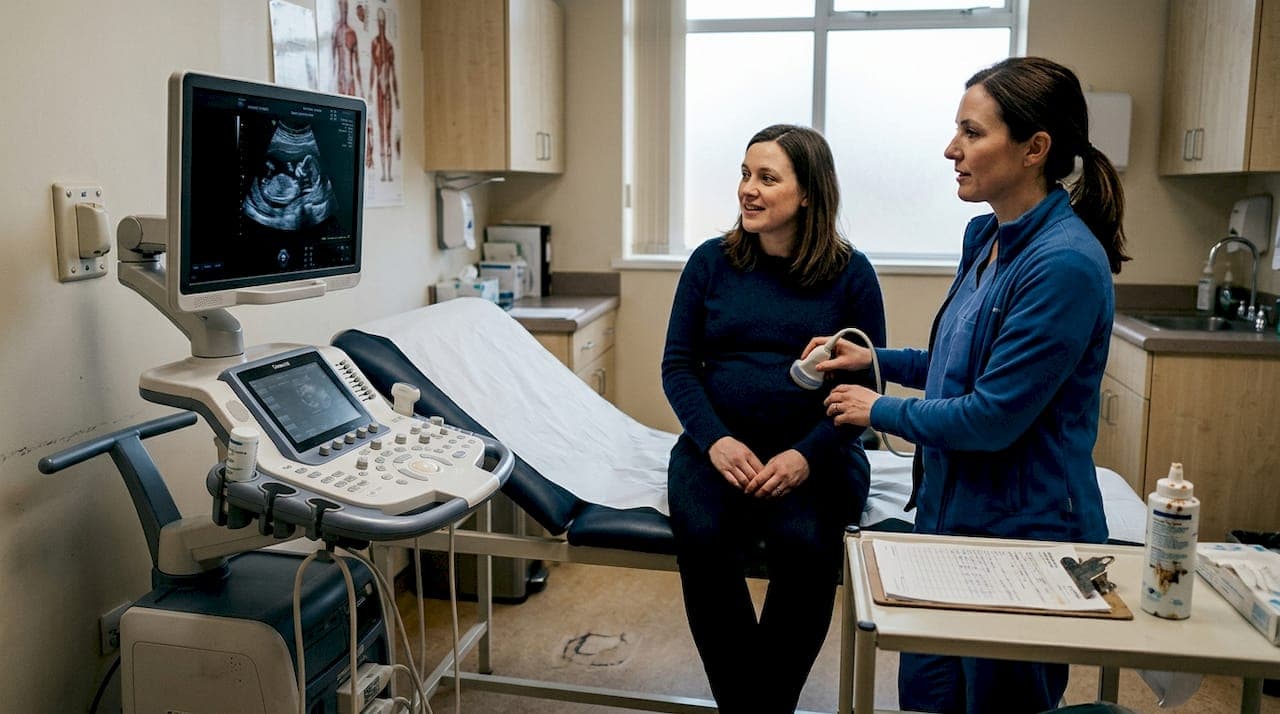
An ultrasound summary is the formal report your provider generates after each scan. It's not just a printout of a blurry image. It's a structured document that records measurements, observations, and clinical findings your care team uses to track your baby's progress throughout pregnancy. Think of it as your baby's first health record.
The technology behind it is straightforward. A transducer device pressed against your skin emits high-frequency sound waves that travel into your body, bounce off fetal tissues, and return as echoes. A computer translates those echoes into the images you see on screen. No radiation involved, just sound.
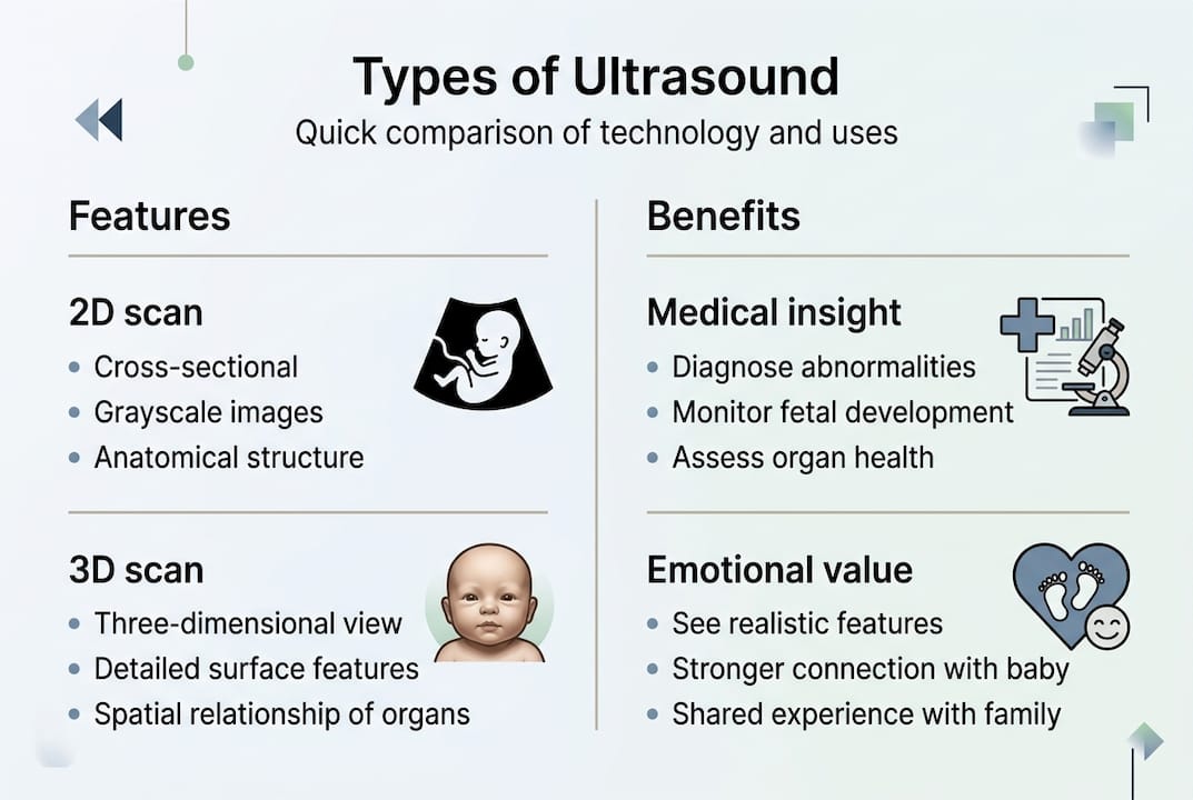
Different imaging modes serve different purposes. B-mode creates 2D images, Doppler measures blood flow velocity through vessels and the umbilical cord, and 3D/4D imaging combines multiple 2D slices to build volume and motion. Each mode adds a layer of clinical or emotional value depending on what your provider needs to assess.
Here's what a standard ultrasound summary typically covers:
- Gestational age based on fetal measurements like crown-rump length or femur length
- Fetal heartbeat and viability confirmation
- Number of fetuses and placental position
- Amniotic fluid levels and uterine anatomy
- Fetal anatomy including brain, spine, heart, kidneys, and limbs
- Growth percentiles compared to expected ranges for gestational age
"The ultrasound summary is your window into your baby's world before birth. Reading it with your provider, not just glancing at it in the parking lot, is where the real value lives."
For parents who want to understand the ultrasound basics before their first appointment, getting familiar with these terms ahead of time makes the conversation with your provider far more productive.
What to expect: Types and timing of prenatal ultrasounds
Understanding the science is key, but most parents are eager to know when scans happen and what each reveals.
Clinical guidelines recommend two routine ultrasounds during a standard pregnancy: a dating scan between 11 and 13 weeks, and an anatomy scan between 18 and 22 weeks. Each serves a distinct purpose.
Here's how the timeline typically unfolds:
- First trimester scan (weeks 6 to 13): Confirms the pregnancy is viable, establishes gestational age, checks for multiples, and screens for early chromosomal markers like nuchal translucency thickness.
- Second trimester anatomy scan (weeks 18 to 22): The most detailed routine scan. Evaluates all major organ systems, checks fetal position, assesses placenta placement, and measures growth against established standards.
- Third trimester growth scans (weeks 28 to 36+): Not always routine but ordered when providers want to monitor growth, fluid levels, or fetal positioning before delivery.
- Advanced elective scans (any trimester, timing varies): 3D and 4D imaging sessions that offer enhanced detail and real-time motion for emotional bonding and, in some cases, better diagnostic clarity.
| Feature | Standard 2D scan | Advanced 3D/4D scan |
|---|---|---|
| Image type | Flat, cross-sectional | Dimensional, surface-rendered |
| Clinical use | Routine diagnostics | Diagnostics plus emotional connection |
| Motion capture | Limited | Real-time with 4D |
| Facial detail | Minimal | High detail |
| Emotional impact | Moderate | Very high |
| Availability | All OB offices | Specialized centers |
Pro Tip: If you're planning an elective 3D/4D session for emotional bonding, the sweet spot is between 26 and 32 weeks. Your baby has enough fat under the skin for clear facial features, but still has room to move freely.
Exploring prenatal ultrasound services that offer advanced imaging options can help you decide which experience fits your pregnancy journey best.
Is ultrasound safe? What the research says
While understanding scan options is exciting, reassurance about safety is just as crucial.
One of the most persistent worries expectant parents carry is whether repeated ultrasound exposure could harm their baby. The short answer: current research says no. Ultrasound does not use ionizing radiation like X-rays or CT scans. It uses sound waves, and that distinction matters enormously.
Key safety facts every parent should know:
- Ultrasound has been used in obstetric care for over 50 years
- No study has confirmed harmful effects on fetal development from clinically indicated scans
- ACOG reports no confirmed risk when ultrasound is used prudently during pregnancy
- The FDA recommends against non-medical "keepsake" ultrasounds performed by untrained operators, not because of sound wave risk, but because of operator qualification concerns
The guiding principle used by sonographers and radiologists is called ALARA, which stands for "As Low As Reasonably Achievable." It means using the minimum exposure time and output needed to get a clear, diagnostically useful image. Certified sonographers are trained to apply this principle on every scan.
Statistic to note: Studies reviewed by major health organizations have consistently found no association between prenatal ultrasound and adverse outcomes including preterm birth, low birth weight, or developmental delays.
Pro Tip: Always ask whether your elective imaging center employs registered diagnostic medical sonographers (RDMS). Certification matters for both safety and image quality.
For more ultrasound safety insights, knowing what questions to ask your provider puts you in a much stronger position during every appointment.
Beyond the basics: What influences summary accuracy and parent experience
After understanding safety, it's time to recognize what shapes the reliability of your ultrasound summary and your overall experience.

Not all ultrasound results are created equal. Several variables affect how accurate and complete your summary will be, and understanding them helps you interpret findings with realistic expectations.
Factors that influence accuracy:
- Operator skill: Sonographer experience directly affects image quality and measurement precision. Accuracy varies significantly based on operator technique, fetal position, and the growth standards used for comparison.
- Fetal position: A baby facing away or curled tightly limits what can be visualized. Providers may ask you to walk, eat something cold, or return for a repeat scan.
- Maternal body composition: Adipose tissue can reduce image clarity, particularly in abdominal scans.
- Equipment quality: Higher-resolution machines produce sharper images and more reliable measurements.
- Gestational age at time of scan: Earlier scans are more precise for dating; later scans carry wider measurement margins.
| Accuracy factor | Impact on summary | How advanced imaging helps |
|---|---|---|
| Operator skill | High | Certified specialists improve precision |
| Fetal position | High | 4D real-time view captures movement |
| Equipment resolution | Moderate to high | HD Live and 8K technology sharpen detail |
| Growth chart standards | Moderate | Customized charts improve LGA detection |
| Gestational age | Moderate | Earlier scans more reliable for dating |
3D and 4D ultrasound is not considered routine medical care, but it offers enhanced emotional value and improved diagnostic clarity in specific cases, particularly for evaluating facial structures, limb anomalies, and fetal behavior.
Beyond the clinical picture, there's something powerful about seeing your baby yawn, stretch, or reach for their face in real time. That moment of recognition, "that's actually my child," is something no report on paper can replicate. Exploring advanced ultrasound options or learning more about BabyView3D can help you understand what's possible beyond the standard clinic experience.
What most parents miss about ultrasound summaries
With these complexities in mind, here's what experience has taught us that typical explanations leave out.
After more than 15 years working with expectant families, we've noticed a pattern. Parents receive their ultrasound summary, scan it for the gender or due date, and tuck it away. The deeper story in that document goes unread.
Borderline findings, measurements at the edge of normal ranges, or notes about fetal position often trigger anxiety without context. But those notations rarely signal a problem. They signal a need for a conversation. Asking your provider "what does this mean for our next steps?" is almost always more useful than searching the term online at midnight.
The other missed opportunity is emotional. Your ultrasound summary is a record of a specific moment in your baby's development. The measurements, the heartbeat rate, the position: these are details you'll want to remember. Pairing that clinical record with a high-quality visual from a professional imaging session creates something genuinely meaningful, not just medically informative.
The parents who get the most from prenatal imaging are the ones who treat it as a collaboration, not a transaction. Ask questions. Request clarification. And give yourself permission to experience the emotional side of it fully.
Discover advanced ultrasound options for your journey
If you want to experience the fullest potential of prenatal imaging, here's where to start.
At BabyView3D, we've spent over 15 years helping families see their babies with stunning clarity through 3D, 4D, and 8K HD Live technology. Our certified sonographers bring both technical precision and genuine warmth to every session.
Whether you're ready to book your first session, explore all ultrasound services available at our centers, or looking to gift a 3D/4D session to someone special, we make it easy to take the next step. Browse our ultrasound photo gallery to see the level of detail that's possible. Your first appointment includes a special introductory offer, because every family deserves to meet their baby before birth.
Frequently asked questions
What key information does an ultrasound summary include?
An ultrasound summary typically covers gestational age, fetal viability, number of fetuses, anatomical development, and placenta location. Reports confirm viability, gestational age, fetal number, anomalies, and placental position as standard components.
How accurate are prenatal ultrasound findings?
Early ultrasounds are highly reliable for establishing gestational age, while later scans carry more variability. Predictive accuracy improves with customized growth charts, though estimates for large-for-gestational-age babies still carry limitations.
Are there any risks to having multiple ultrasounds during pregnancy?
Current research shows no confirmed risks when ultrasounds are used as clinically indicated. No risk is associated with prenatal ultrasound when performed prudently, according to ACOG guidelines.
What is the difference between 2D, 3D, and 4D ultrasounds?
2D is the standard for medical diagnostics, 3D adds surface depth and facial detail, and 4D captures real-time motion. 3D/4D provides volume and motion imaging but is not considered routine care and is primarily used to enhance the parental bonding experience.