Most expecting parents use the words "ultrasound" and "sonogram" like they mean the same thing. They're related, but there's an important difference, and once you understand it, everything about your prenatal appointments starts to feel clearer. A sonogram is the image produced during an ultrasound exam, not the exam itself. This guide walks you through how sonograms work, what the different types show, what science says about safety, and how to make each appointment a meaningful moment you'll remember long after your baby arrives.
Table of Contents
- What is a sonogram? The basics explained
- The different types of sonograms in pregnancy
- Is it safe? What science says about sonogram safety
- Sonogram experiences: What to expect and how to make the most of them
- From our perspective: Sonograms are more than just pictures
- Experience your sonogram with BabyView3D
- Frequently asked questions
Key Takeaways
| Point | Details |
|---|---|
| Sonogram definition | A sonogram is the picture created during an ultrasound exam, showing your developing baby. |
| Variety of types | Pregnancy sonograms include 2D, 3D, 4D, and Doppler, each serving a unique purpose. |
| Safety backed by science | Routine sonograms are safe when performed by professionals, following strict guidelines. |
| Beyond medical, it's emotional | Sonograms also foster memorable moments and connections for parents with their babies. |
What is a sonogram? The basics explained
Let's start with a definition that actually sticks. An ultrasound is the medical procedure. A sonogram is the picture that comes out of it. Think of it this way: an ultrasound is the camera, and a sonogram is the photograph. The terms are used interchangeably in everyday conversation, but knowing the distinction helps you understand exactly what your provider is doing and why.
The technology behind sonograms is genuinely fascinating. High-frequency sound waves between 2 and 18 MHz are sent from a small device called a transducer probe into your body. Those waves bounce off tissues, organs, and your growing baby, creating echoes that a computer processes into visual images. No radiation. No needles. Just sound waves and math.
Those images can take several forms:
- 2D sonograms: The classic flat, gray-scale image most people picture
- 3D sonograms: A still, three-dimensional surface image of your baby's features
- 4D sonograms: A live, moving version of the 3D image, like a short video
- HD Live: Ultra-realistic imaging that adds lighting effects for lifelike detail
Sonograms can show fetal position, the size and growth of your baby, the location of the placenta, amniotic fluid levels, and whether your baby's organs are developing normally. Each image tells a story about your pregnancy. You can explore our sonogram imaging services to see what's possible at different stages.
"The ultrasound exam is one of the most important tools in prenatal care, giving providers a real-time window into fetal development without any invasive procedures."
Pro Tip: During your next appointment, ask your sonographer to narrate what they're seeing in real time. You'll walk away with a much richer understanding of those images, and the experience becomes instantly more memorable.
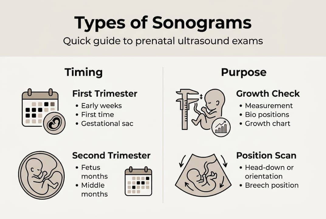
The different types of sonograms in pregnancy
Not every sonogram is the same. Different types serve different purposes, and knowing what each one involves helps you know what to expect when your provider recommends one.
Here's a quick breakdown of the most common types:
- Transabdominal: A probe glides over your belly. This is the standard method used in most second and third trimester checkups.
- Transvaginal: A small probe is inserted internally. It's used in early pregnancy when the baby is too small for a clear abdominal view.
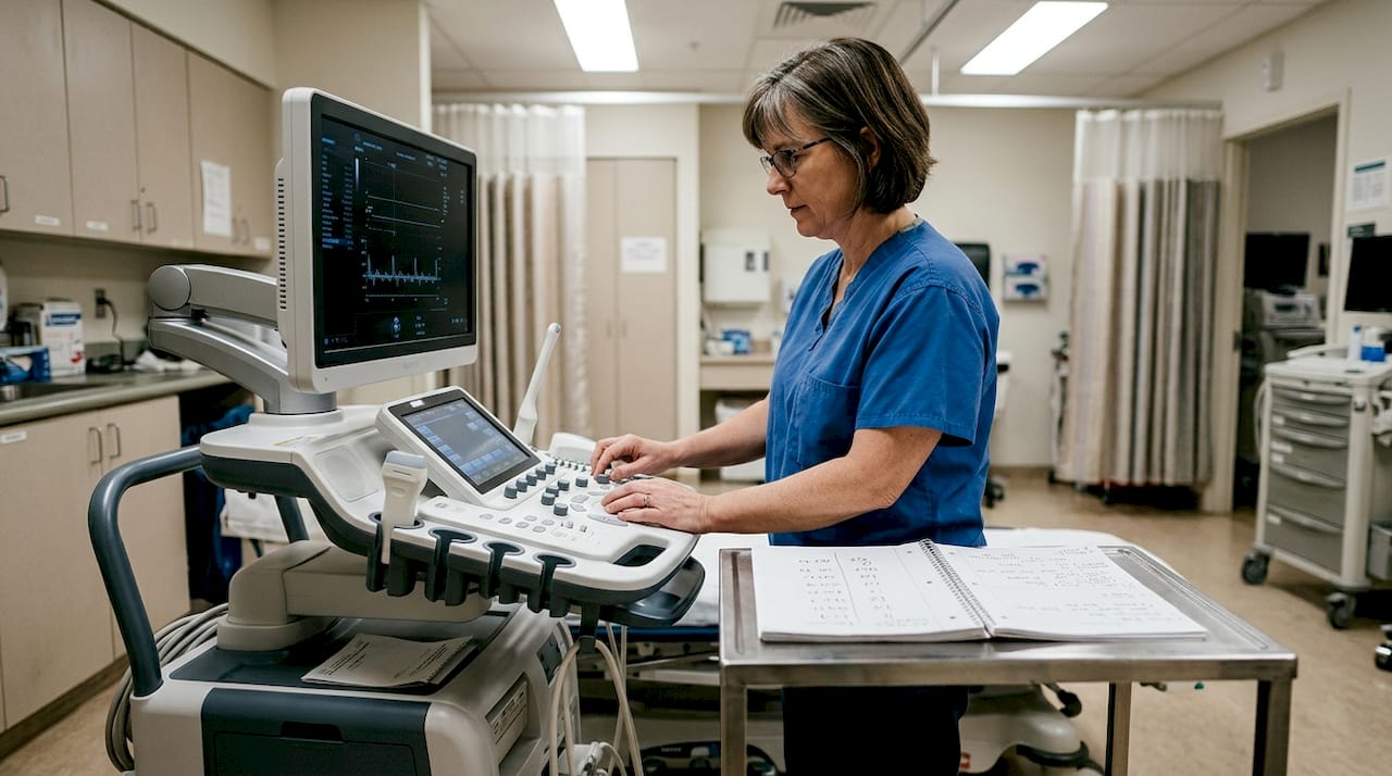
- Doppler: Measures blood flow through the umbilical cord and fetal heart. Critical for monitoring circulation and detecting any concerns.
- 3D and 4D: Surface imaging that captures detailed views of your baby's face and movements. More emotionally vivid than standard imaging.
Timing matters too. Routine ultrasounds are offered at 11 to 14 weeks for dating and nuchal translucency screening, and again between 18 and 21 weeks for the anatomy scan.
| Sonogram type | When it's used | What it shows |
|---|---|---|
| Transabdominal | 12 weeks onward | Growth, position, placenta |
| Transvaginal | 6 to 12 weeks | Early viability, heartbeat |
| Doppler | Any trimester | Blood flow, circulation |
| 3D | 26 to 32 weeks | Surface detail, facial features |
| 4D | 26 to 32 weeks | Real-time movements, bonding |
| HD Live | 26 to 32 weeks | Photorealistic imaging |
For a deeper look at what each procedure involves, check out the detailed sonogram procedures available through our resource center.
Pro Tip: While 3D and 4D sonograms create incredible bonding experiences, the FDA and ACOG advise against non-medical use purely for keepsake photos. Always choose a provider who follows established safety protocols and has certified sonographers on staff.
Is it safe? What science says about sonogram safety
This is the question almost every parent asks, and the answer is reassuring. Decades of research support the safety of diagnostic prenatal ultrasound when it's performed correctly and with clinical purpose.
The FDA sets strict output limits for diagnostic equipment. Mechanical Index must stay at or below 1.9, and spatial peak temporal average intensity must not exceed 720 mW per cm² for standard diagnostic use. Ophthalmic exams have an even tighter limit of 0.23. These thresholds exist to protect you and your baby.
Every properly trained sonographer also follows the ALARA principle, which stands for "As Low As Reasonably Achievable." It means they always use the lowest possible output for the shortest necessary time to get a clear image. Safety is not an afterthought. It's built into the process.
The research behind these guidelines is solid. No confirmed adverse effects have been found from diagnostic prenatal ultrasound in human studies. Some animal studies at unusually high exposure levels have raised theoretical concerns, but those conditions don't reflect real clinical practice. The overwhelming consensus from the medical community is that routine prenatal sonograms are safe.
"When performed by trained professionals following established guidelines, diagnostic ultrasound poses no confirmed risk to mother or baby. It remains one of the most valuable and safe tools in prenatal medicine."
Key safety points to keep in mind:
- Only use providers with certified sonographers
- Avoid purely recreational, non-medical sessions at unregulated facilities
- Ask your provider about their equipment and safety standards if you have concerns
- First trimester scans use B-mode or M-mode, which carry the lowest risk profile
We go deeper on the research behind prenatal ultrasound safety if you'd like to explore the evidence for yourself.
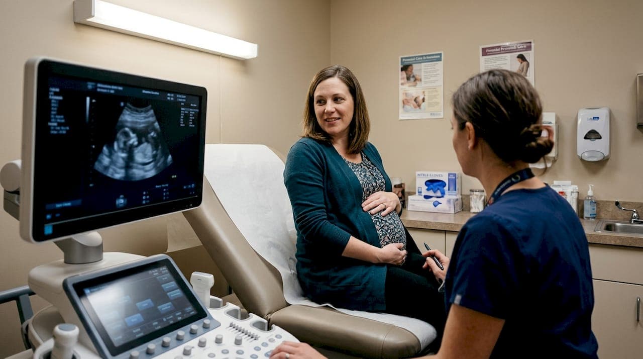
Sonogram experiences: What to expect and how to make the most of them
Knowing that sonograms are safe is one thing. Knowing how to actually show up for the experience is another. The more prepared you are, the more meaningful each appointment becomes.
Here's what a typical sonogram appointment looks like, step by step:
- Arrive hydrated. For transabdominal scans, a full bladder helps produce a clearer image. Your provider will usually give specific instructions beforehand.
- Check in and get settled. Sessions typically run 10 to 45 minutes depending on the type of scan and what your provider needs to assess.
- Gel is applied to your skin. It looks cold and odd, but it helps the transducer make full contact for better sound wave transmission.
- The scan begins. Your sonographer moves the probe and captures images from different angles. They may go quiet during measurement, so don't worry if it gets silent.
- Results are discussed. Your provider explains what they saw. This is the time to ask questions, not after you leave.
- Ask about keepsakes. Many providers offer printed or digital images. Some offer short video clips of 4D sessions.
One thing worth understanding: sonogram images have limits. A gestational sac over 25mm with no embryo, or a crown-rump length above 7mm with no heartbeat, would signal a non-viable pregnancy. These edge cases are why trained interpretation matters so much.
You can browse our prenatal sonogram packages to compare options that fit your stage of pregnancy and your goals for the experience.
Pro Tip: Bring your partner or a support person. Ask the sonographer to explain every measurement they take in plain language. And consider requesting a short video clip if your provider offers 4D. That footage becomes something you'll watch for years.
From our perspective: Sonograms are more than just pictures
We've worked with families for over 15 years, and one pattern shows up again and again: parents who treat the sonogram appointment as purely routine miss something irreplaceable. They get the image. They glance at it. They move on. And that's a genuine loss.
A sonogram is not just a diagnostic step. It's often the first time a family truly sees their baby as a person. A nose. A yawn. A tiny hand raised near a cheek. Those details don't just confirm a healthy pregnancy; they begin a relationship. That's not sentimental fluff. That's exactly what the experience is capable of delivering when parents choose to engage with it fully.
The mistake we see most often is waiting passively for the "photo op" instead of being present for the whole experience. Ask questions. Watch the screen. Let yourself feel the weight of what you're looking at. The sonogram stories that stay with families forever are the ones where parents were fully there, not just physically but emotionally.
Technology matters, absolutely. Better imaging means more detail, more clarity, more chances to truly see your baby before birth. But the technology only serves you when you bring your full attention to the moment it creates.
Experience your sonogram with BabyView3D
If you want a sonogram experience that goes beyond a routine checkup, we built BabyView3D for exactly that. Our certified sonographers use the latest 3D, 4D, and 8K HD Live technology to give you the clearest, most emotionally powerful view of your baby possible.
Exploring your sonogram service options takes just a few minutes, and you can start to understand exactly what each package offers for your stage of pregnancy. Want to see what your experience could look like before you book? Browse our sonogram photo gallery to see real images from real families. We have locations across the United States, first-appointment discounts available, and a team that's genuinely invested in making this a moment you'll never forget.
Frequently asked questions
What is the difference between an ultrasound and a sonogram?
An ultrasound is the exam or technology used, while a sonogram is the image produced during that exam. Most people use the terms interchangeably, but technically they describe two different things.
Are 3D or 4D sonograms safe for my baby?
Yes, when performed by trained professionals following medical guidelines, 3D and 4D sonograms are considered safe. However, non-medical elective use is not recommended by the FDA or ACOG due to unnecessary exposure without clinical purpose.
How many sonograms will I have during a normal pregnancy?
Most standard pregnancies include two routine ultrasounds: one between 11 and 14 weeks for dating and nuchal screening, and another between 18 and 21 weeks for the anatomy scan. High-risk pregnancies may involve more frequent monitoring.
Can I keep a copy of my baby's sonogram?
Yes. Most providers offer printed sonogram images or digital copies to take home, and some offer video clips from 4D sessions as keepsakes you can share with family.
Is there a risk to having extra or elective sonograms?
Unnecessary non-medical ultrasounds should be avoided to minimize potential risk. The FDA and ACOG specifically caution against elective non-medical sessions at facilities without proper clinical oversight.