Not all ultrasound sessions are the same experience, and understanding the difference can completely change how you prepare for each appointment. Some parents walk in expecting a magical, crystal-clear view of their baby's face, only to find themselves staring at a blurry black-and-white image on a small screen. Others are surprised to learn that advanced 3D and 4D sessions exist specifically for bonding and memory-making. Knowing what each type of session offers, what to realistically expect, and how to get the most out of every scan helps you show up informed, emotionally ready, and fully present for one of the most meaningful chapters of your life.
Table of Contents
- What is an ultrasound session?
- Routine ultrasound milestones in prenatal care
- Medical, detailed, and keepsake ultrasounds: What's different?
- Understanding the quality and limits of ultrasound exams
- What expectant parents miss about ultrasound sessions
- Explore advanced ultrasound experiences with BabyView3D
- Frequently asked questions
Key Takeaways
| Point | Details |
|---|---|
| Not all sessions are the same | Ultrasound sessions vary by purpose, from medical diagnosis to emotional bonding opportunities. |
| Key milestones | Most parents will have two essential scans—the dating and anatomy ultrasounds—during pregnancy. |
| Expect incomplete results | A scan may not capture everything in one session due to normal factors; repeat scans are common. |
| Understand safety principles | Ultrasounds are safe when guided by professional standards emphasizing minimal and prudent use. |
What is an ultrasound session?
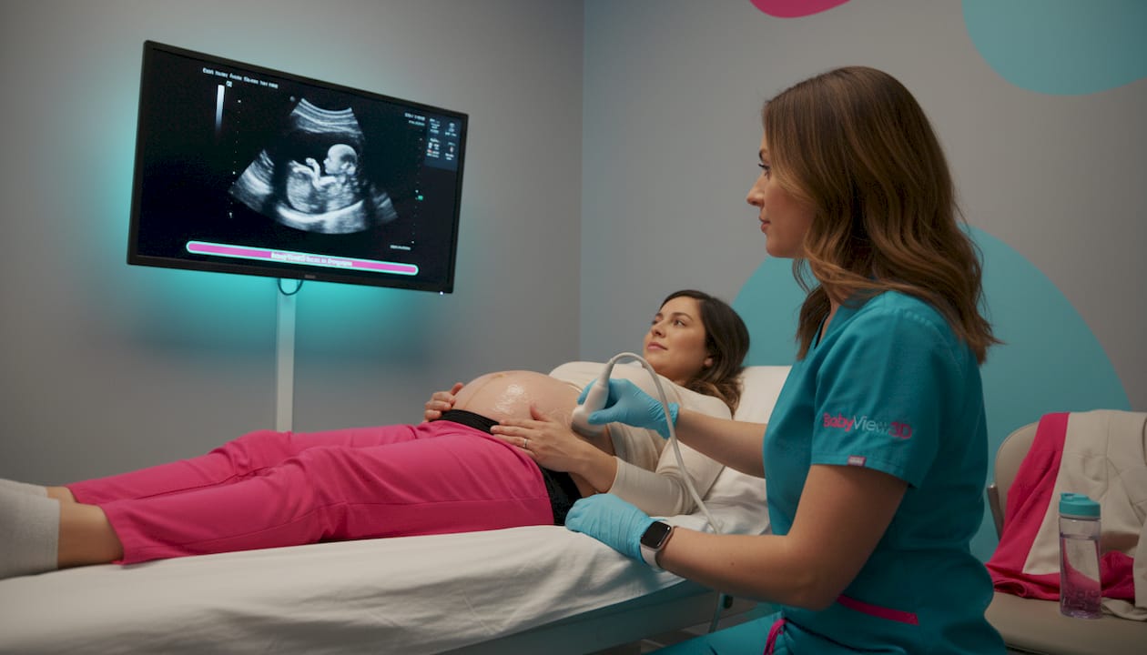
At its core, an ultrasound session is a scheduled appointment where a sonographer or medical provider uses high-frequency sound waves to create real-time images of your developing baby. The equipment sends sound waves into your body, which bounce off tissues and return as signals that are translated into visual images. No radiation is involved, which is a key reason ultrasound is the standard imaging choice throughout pregnancy.
But what happens in that room goes well beyond a simple medical checkup. Ultrasound sessions serve two broad purposes. The first is clinical: tracking fetal development, confirming pregnancy location, assessing placenta position, and identifying any concerns that need attention. The second is deeply personal: giving you and your family a real, visual connection to the baby growing inside you. These two goals often coexist, but they are not always the same kind of session.
Here is a quick look at the types of scans you may encounter:
- 2D ultrasound: The classic black-and-white image most people associate with prenatal scans. Standard in clinical settings.
- 3D ultrasound: Produces a still three-dimensional image of your baby's face and features with remarkable detail.
- 4D ultrasound: The same as 3D, but in real-time video. You can watch your baby yawn, stretch, or suck their thumb.
- HD Live / 8K ultrasound: The most advanced imaging available today, producing lifelike images with depth, shadow, and stunning clarity.
"Ultrasound is the imaging technique of choice in pregnancy and should be used prudently for clinical benefit." — ACOG Committee Opinion No. 723
Safety has always been a central conversation around ultrasound. Medical organizations affirm that ultrasound is not associated with known fetal harm when used appropriately. The key phrase is "used prudently." This means sessions should have a clear purpose, whether that is medical evaluation or an intentional, supervised keepsake experience. Understanding when ultrasound sessions are recommended can help you make informed decisions about how many sessions are right for your pregnancy.
Pro Tip: Write down your questions before every appointment. A prepared parent gets more out of every session, whether it is a routine check or a special keepsake scan.
Routine ultrasound milestones in prenatal care
Now that we know what an ultrasound session is, let's break down the typical journey through prenatal ultrasound milestones. Most U.S. parents will experience two major scheduled ultrasounds, though additional scans are often ordered based on individual clinical circumstances.
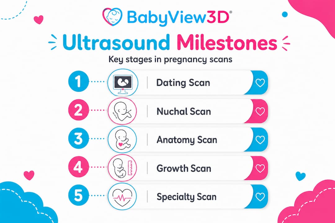
Here is a numbered overview of the standard prenatal ultrasound timeline:
- First trimester dating scan (6–12 weeks): Confirms the pregnancy, establishes gestational age, checks for a heartbeat, and rules out ectopic pregnancy (a pregnancy that develops outside the uterus). This is often the first time you hear that fast, rhythmic sound that makes everything feel real.
- Nuchal translucency scan (11–14 weeks): Measures fluid at the back of the baby's neck to screen for chromosomal conditions. Not universally ordered, but commonly offered.
- Anatomy scan (18–22 weeks): The most detailed routine scan. A trained sonographer checks every major organ system, limbs, the placenta, and amniotic fluid. This is also often when families find out the baby's sex.
- Third trimester growth scan (28–36 weeks): Sometimes ordered when the provider wants to monitor growth, fluid levels, or placental function more closely.
- Late-term biophysical profile: Evaluates fetal movement, breathing, tone, and fluid. Typically only ordered when there is a specific clinical concern.
| Scan type | Typical timing | Primary purpose |
|---|---|---|
| Dating scan | 6–12 weeks | Confirm pregnancy, establish due date |
| Nuchal translucency | 11–14 weeks | Screen for chromosomal conditions |
| Anatomy scan | 18–22 weeks | Detailed structural assessment |
| Growth scan | 28–36 weeks | Monitor size and fluid |
| Biophysical profile | 36+ weeks | Assess fetal well-being |
According to prenatal care guidelines, routine prenatal care includes a first-trimester scan and a detailed anatomy scan in the second trimester, with timing typically around 12 and 18–22 weeks. Most parents have at least these two scans. Everything beyond that is driven by clinical need or personal choice. Having a clear overview of ultrasound timelines helps you ask smarter questions at each appointment and understand what your provider is looking for.
Key statistic: The anatomy scan is performed between 18 and 22 weeks because this is when fetal structures are large enough to assess clearly but the baby is still positioned in a way that allows a full view. Timing matters significantly for scan completeness.
Medical, detailed, and keepsake ultrasounds: What's different?
Having covered routine milestones, it is important to understand how different types of specialized ultrasounds compare and what to look for when considering your options.
Not every ultrasound appointment has the same goal. A medical ultrasound conducted by your OB or midwife is focused entirely on clinical data. A detailed anatomical ultrasound, often performed by a maternal-fetal medicine specialist (a perinatologist), takes that further and examines fetal structures with significantly more depth and time. A keepsake or bonding session, like those offered at dedicated imaging studios, shifts the focus entirely to the emotional experience of meeting your baby before birth.
Here is a side-by-side comparison to clarify the differences:
| Feature | Medical ultrasound | Detailed/specialist exam | Keepsake/bonding session |
|---|---|---|---|
| Primary goal | Clinical monitoring | Comprehensive structural review | Emotional connection and memory-making |
| Technology | Standard 2D/Doppler | High-resolution 2D/3D | 3D, 4D, HD Live, or 8K |
| Performed by | OB, midwife, tech | Perinatologist or specialist | Certified sonographer |
| Insurance coverage | Usually covered | Often partially covered | Generally not covered |
| Appointment length | 20–45 minutes | 45–90 minutes | 30–60 minutes |
| Keepsakes (photos/video) | Occasionally | Rarely | Central part of the experience |
What you should know before booking any session beyond your standard care:
- Ask whether the facility follows the ALARA principle (As Low As Reasonably Achievable), which guides safe exposure levels during scanning.
- Confirm that staff are certified sonographers with documented training and experience in prenatal imaging.
- Understand that keepsake ultrasound sessions are marketed for bonding but medically should follow the ALARA principle and emphasize prudent use.
- Check whether the studio uses up-to-date equipment and follows hygiene and safety protocols.
Pro Tip: Before booking a keepsake session, ask the studio directly about their sonographers' credentials, their safety protocols, and how long their sessions typically run. A reputable provider welcomes these questions.
Exploring your ultrasound options and services before committing gives you the confidence to walk into that room knowing what to expect and who is caring for you and your baby.
Understanding the quality and limits of ultrasound exams
Now that we have compared types of ultrasounds, it is important to set real expectations about how thorough scans can be in practice. Even the most advanced equipment cannot guarantee a complete, clear view of every structure in every session.
The completeness of an ultrasound exam depends on several factors that are largely outside anyone's control. Baby's position is the biggest variable. If your little one has their back turned, face buried, or legs crossed, some structures simply cannot be imaged properly. Maternal anatomy, including body composition, scar tissue from previous surgeries, or the position of the uterus, can also affect image clarity.
Here is a data snapshot from recent medical research:
| Study variable | Finding |
|---|---|
| Range of incomplete exam rates | Less than 4% to 57% depending on facility and population |
| Most common cause | Fetal position at time of scan |
| Resolution rate | Most incomplete scans are resolved with a repeat appointment |
That wide range, less than 4% to more than half in some settings, reflects how dramatically facility quality, sonographer experience, and patient factors can influence outcomes. This is not a reason to panic. It is a reason to choose your imaging provider carefully.
Research on detailed ultrasound quality shows that incomplete exam rates vary widely, meaning repeat scans are sometimes a normal and necessary part of the process, not a sign of a problem.
Common reasons a scan may be incomplete or require a repeat visit:
- Baby is in a position that blocks key views (especially common during the anatomy scan window)
- Low amniotic fluid makes imaging more difficult
- Baby is moving too actively for still measurements
- Maternal body composition affects ultrasound wave penetration
- Equipment limitations at certain facilities
What can you do to improve your chances of a clear, complete session? Follow any instructions your provider gives about hydration or food intake beforehand. Schedule your appointment at a time when you know your baby tends to be more active. And if you are going for a keepsake session, choose a provider with advanced technology and experienced staff. How detailed exams are assessed matters enormously, and facilities that prioritize image quality consistently show better results.
What expectant parents miss about ultrasound sessions
Here is a perspective that comes from years of working closely with families through prenatal imaging experiences: most parents focus so intensely on what they hope to see that they forget to prepare for what they might not.
The emotional weight of an ultrasound appointment is real and significant. You are going to see your baby. You might watch them kick or yawn. You might finally get a clear view of their face. That anticipation is beautiful, and it deserves to be honored. But there is a side of the experience that no one really prepares you for.
Sometimes the baby does not cooperate. Sometimes the images are fuzzy. Sometimes the sonographer needs to stay quiet because they are concentrating on clinical measurements, and the room feels unexpectedly tense. Sometimes a repeat visit is needed, and parents leave feeling deflated when they were expecting to feel overjoyed.
What we have learned from over 15 years of working with families is this: the parents who leave most satisfied are the ones who came with flexible expectations and genuine curiosity, not a rigid script for how the session should go. They asked questions. They stayed present. They understood that an incomplete view today does not close the door on connection.
The clinical and emotional sides of prenatal care do not have to compete with each other. A routine scan can be a deeply moving moment if you let it be. A keepsake session can feel hollow if you come in hoping it will replace information you did not get at your medical appointment. Balance and preparation are everything. Following ultrasound best practices going into each session helps you protect both your peace of mind and your ability to be present in that room.
Pro Tip: Set a flexible intention before each session. Instead of "I need to see the baby's face clearly," try "I'm here to connect with my baby and support their health." This small mental shift can make the entire experience more rewarding, regardless of what the screen shows.
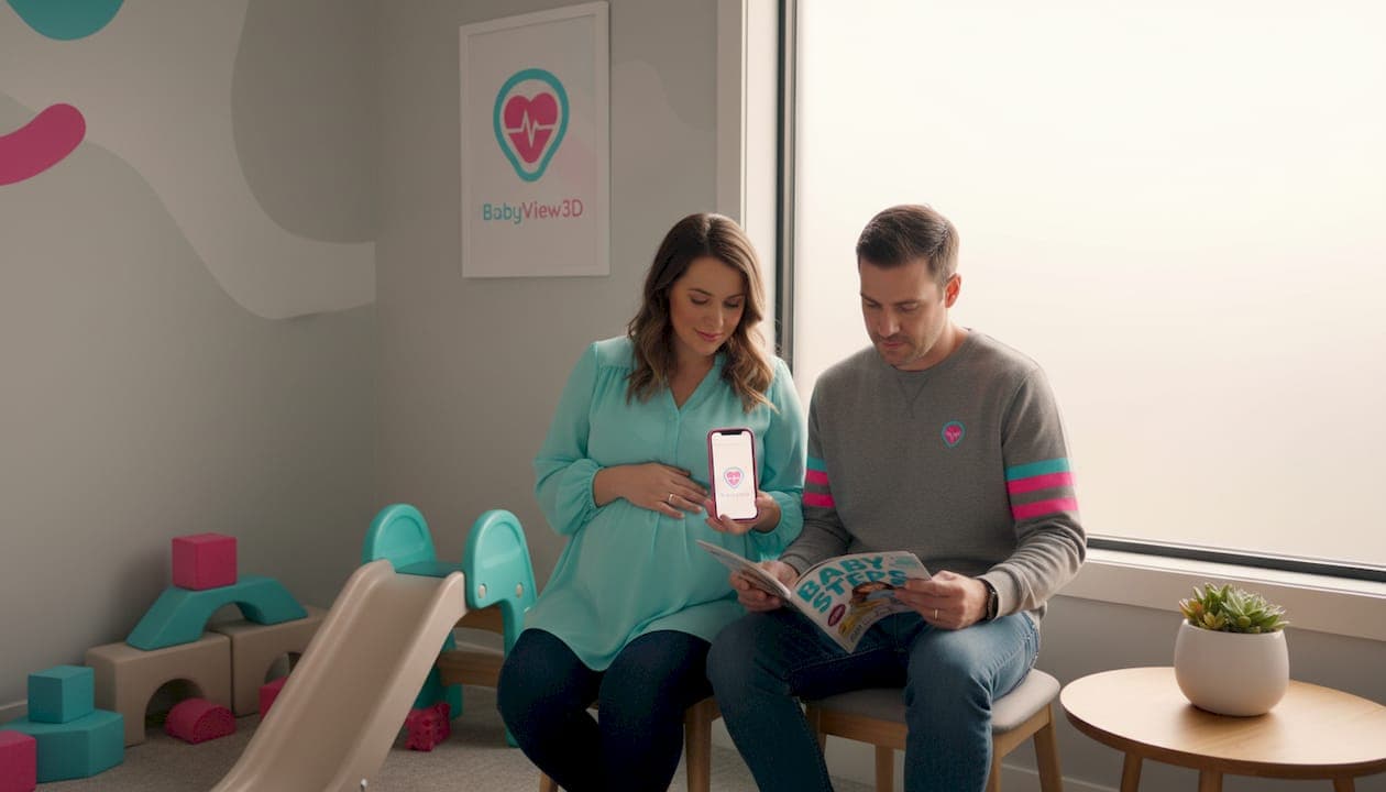
Explore advanced ultrasound experiences with BabyView3D
If you are ready to apply what you have learned, BabyView3D is here to make your next ultrasound session truly special.
BabyView3D offers a range of ultrasound options designed for families who want both safety and stunning image quality. With over 15 years of experience, certified sonographers, and the most advanced 3D, 4D, and 8K HD Live technology available in the U.S., every session is crafted around your family's emotional experience and peace of mind. Whether you want a first-trimester peek, a detailed bonding session mid-pregnancy, or a final memorable visit before your baby arrives, there is a package for every stage. Browse real session memories from families who have shared their experience with us, and book your first appointment with a special first-visit offer.
Frequently asked questions
How many ultrasound sessions are typical during pregnancy?
Most parents in the United States have at least two ultrasounds: a first-trimester dating scan and a second-trimester anatomy scan. Routine prenatal care includes both a first and second trimester ultrasound session, with additional scans ordered based on clinical need.
Are 3D or 4D ultrasounds safe for my baby?
When performed by trained professionals following medical guidelines, 3D and 4D ultrasounds are safe but should be used with purpose. Ultrasonography is not associated with risk but should be used prudently throughout pregnancy.
Why might my anatomy ultrasound session be incomplete?
Incomplete sessions often result from baby's position, maternal anatomy, or technical limits, with repeat scans sometimes needed for a full assessment. Incomplete rates for detailed anatomy scans can range widely based on several factors, and a repeat visit is a normal part of the process.
Can I bring family or record my ultrasound session?
Many facilities welcome family and allow recording parts of the session, but it is best to check your provider's specific policies before your appointment, especially for medical scans.
Do insurance plans cover keepsake ultrasounds?
Keepsake and bonding ultrasounds that are not medically necessary are generally not covered by insurance. Budget for these sessions separately and treat them as the premium, memorable experience they are designed to be.