

Ultrasound in pregnancy is one of the most significant medical tools your care team has, yet most expectant parents walk into their first scan thinking it is simply a chance to see their baby. It is that, but it is also far more. A pregnancy ultrasound is a non-invasive, painless imaging test that uses high-frequency sound waves to generate images of your developing baby, your placenta, and your uterus. It carries no radiation and has no known side effects. Understanding what it actually does will help you prepare better, ask smarter questions, and approach every appointment with both excitement and clarity.
Table of Contents
- Key Takeaways
- What is ultrasound in pregnancy: how it works and why it's safe
- Types of pregnancy ultrasound scans and when you'll have them
- The role of the sonographer: more than a technician
- What to expect at your ultrasound appointment
- My perspective: what families consistently underestimate
- See your baby with confidence at Bbview3d
- FAQ
Key Takeaways
| Point | Details |
|---|---|
| Ultrasound uses sound, not radiation | High-frequency sound waves create images with no known risks to you or your baby. |
| Timing of scans matters | The first trimester scan and the 18 to 22 week anatomy scan serve very different medical purposes. |
| Sonographers are skilled professionals | They detect abnormalities, assess growth, and guide families through emotionally complex moments. |
| Preparation improves your scan | Knowing what to bring, how to hydrate, and what to expect reduces stress and improves image quality. |
| Ultrasound is a medical exam | Scans are often joyful, but families should be ready for the full range of possible findings. |
What is ultrasound in pregnancy: how it works and why it's safe
At its core, a pregnancy ultrasound works like a very precise form of sonar. A handheld device called a transducer sends high-frequency sound waves into your body. Those waves bounce off internal structures including the baby, the uterus, and the placenta, then return to the transducer. A computer translates the echoes into real-time images on a screen. The whole process happens in seconds, continuously, which is why you can see your baby moving during the scan.
What makes ultrasound particularly well suited for pregnancy is what it does not use. Unlike X-rays or CT scans, ultrasound produces no ionizing radiation, which means there is no cumulative exposure risk for you or your developing baby. Decades of research and clinical use confirm its safety profile. It remains the preferred imaging method throughout all stages of pregnancy for exactly this reason.
The role of ultrasound in prenatal care goes well beyond confirming a pregnancy. Here is what a standard ultrasound assessment covers:
- Fetal growth monitoring: Measuring the baby's head, abdomen, and femur length to track development against expected ranges
- Placenta position: Identifying whether the placenta is positioned in a way that could complicate delivery
- Amniotic fluid levels: Checking that there is enough fluid to support fetal movement and organ development
- Fetal heartbeat: Confirming cardiac activity, particularly in the first trimester
- Structural assessment: Evaluating the baby's organs, spine, and limbs for visible abnormalities
- Cervical length: Measuring the cervix to assess preterm birth risk in some cases
One important point about the limits of ultrasound: it is excellent at detecting structural issues and tracking growth, but it cannot see everything. Chromosome conditions such as Down syndrome require additional blood tests or diagnostic procedures. Ultrasound is one layer of prenatal care, not the complete picture.
Types of pregnancy ultrasound scans and when you'll have them
Not all pregnancy ultrasounds serve the same purpose. The type of scan you receive depends on your gestational age, your pregnancy risk profile, and what your care provider needs to assess. Timing is not arbitrary. Each scan is scheduled to capture information that is only visible or measurable during a specific window of fetal development.
| Scan type | Timing | Primary purpose |
|---|---|---|
| Dating or viability scan | 6 to 10 weeks | Confirms heartbeat, estimates due date, detects multiples |
| First trimester screen | 11 to 13 weeks | Measures nuchal translucency, screens for chromosomal risk |
| Anatomy scan | 18 to 22 weeks | Full structural assessment of fetal organs, spine, and limbs |
| Growth scan | 28 to 36 weeks | Monitors growth rate, fluid levels, and placenta function |
| Biophysical profile | Third trimester | Assesses fetal tone, breathing movements, and fluid in high-risk cases |
The WHO recommends at least one ultrasound before 24 weeks for every pregnant woman to estimate gestational age, identify multiple pregnancies, and detect major anomalies. Many care providers in the United States schedule two or more standard scans, with additional scans for pregnancies considered higher risk.
The anatomy scan, which typically happens between weeks 18 and 22, is the most detailed routine scan you will have. It lasts between 30 and 90 minutes and systematically evaluates the baby's brain, heart, kidneys, spine, limbs, face, and abdominal organs. Research shows it can detect approximately 50 percent of major structural malformations. Parents with higher BMIs may require follow-up scans because abdominal tissue can reduce image clarity.

First trimester scans carry their own emotional weight. Hearing a heartbeat for the first time or learning you are carrying twins creates a moment that stays with families forever. Those early scans also give your provider the data needed to calculate your due date accurately, which affects every clinical decision from that point forward.
The role of the sonographer: more than a technician
The person operating the ultrasound machine carries more responsibility than most expectant parents realize. Sonographers are skilled healthcare professionals trained to assess fetal growth, identify abnormalities, and communicate findings to both physicians and families. Their work sits at the intersection of clinical precision and human sensitivity.
Technically, a sonographer must capture dozens of standardized measurements and images within a single appointment, often while managing a fetal subject who moves unpredictably. Getting a clear view of the four chambers of the heart or the position of the spine requires expertise that comes from years of training and practice.
"Sonographers act not just as technicians but as patient advocates who balance empathy with technical proficiency at every exam." — Sonographer Salary
The emotional layer of this role is just as demanding. A sonographer who spots a potential abnormality cannot simply announce it in the room. They are trained to complete the exam professionally, maintain composure, and ensure the right clinical team delivers and interprets findings properly. That restraint protects families from premature alarm while ensuring the right follow-up happens.
Advanced practice sonographers with credentials like RDMS can identify subtle abnormalities missed during routine exams. Research suggests these specialists identify issues in up to seven percent of ultrasounds initially deemed normal. The expertise level of your sonographer genuinely affects outcomes.
Pro Tip: When you schedule your anatomy scan, ask whether the facility employs credentialed diagnostic medical sonographers. Certification matters more than the technology in the room.
What to expect at your ultrasound appointment
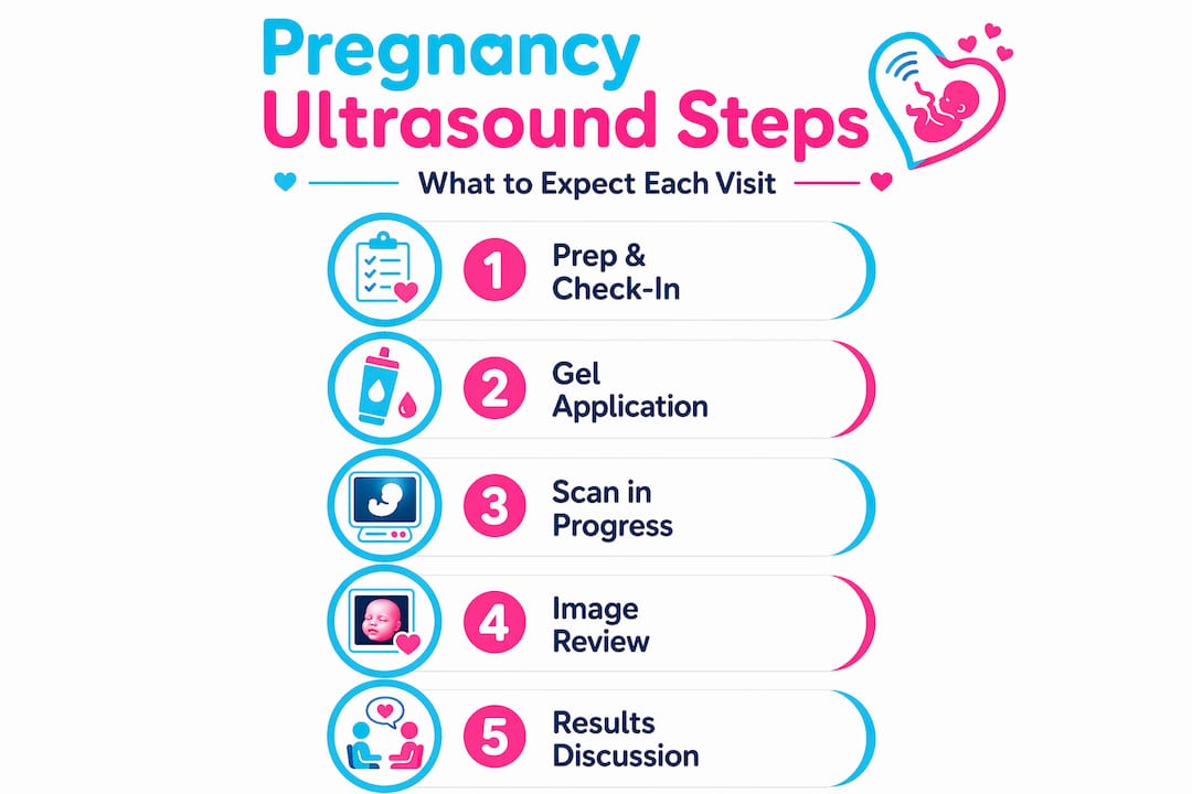
Walking into an ultrasound appointment prepared makes the whole experience less stressful and often produces better images. Here is a step-by-step picture of how a standard appointment unfolds.
- Check-in and preparation. You will provide your pregnancy dates and any relevant history. Your sonographer will explain the process before beginning.
- Positioning. You will lie on an exam table. For most scans, a gel is applied to your abdomen and the transducer is moved across your skin.
- Image capture. The sonographer works systematically through a checklist of measurements and views. This is not a casual browse. Every image serves a clinical purpose.
- Bladder management. For some scans, a full bladder improves image quality by pushing the uterus into a better position. For others, you may be asked to empty your bladder mid-exam or have a transvaginal scan to get clearer views of the cervix or placenta.
- Fetal repositioning. Babies do not cooperate on command. Your sonographer may ask you to walk around, shift position, or wait while the fetus moves into a better angle for imaging.
- Review. Once the required images are captured, your provider or a physician will review results. Some findings are shared immediately. Others require further evaluation.
One thing that surprises many parents: photography and video recording are typically prohibited during diagnostic scans. Additional lighting from phone screens can distort images on the monitor, and sonographers need full concentration. You will receive printed images or a digital summary after the exam.
Emotionally, the most important thing you can do is walk in prepared for the full range of outcomes. Ultrasound scans are medical evaluations, not performances. Most scans deliver joyful news, but some reveal concerns that require follow-up. Knowing this in advance does not dampen the joy of a healthy result. It simply means you are going in as an informed, prepared parent rather than one caught off guard.
Pro Tip: Bring a support person who can focus on the emotional experience while you focus on listening to the sonographer. Two sets of ears always catch more information.
My perspective: what families consistently underestimate
I have spent years immersed in the world of prenatal imaging, and what I keep seeing is the same gap. Families come in expecting a preview. They leave understanding they just had a medical exam. The emotional shift in the room when something unexpected appears on the screen is something I will never forget witnessing.
What I have learned is that the sonographer in that room is doing two jobs simultaneously. They are building a precise clinical record while also managing the emotional reality of a family watching their baby on a screen for the first time. That dual responsibility is exhausting and underappreciated.
I have also noticed that the families who handle unexpected findings best are the ones who came in having already asked themselves: "What if this scan shows something we didn't expect?" That question is not pessimistic. It is preparation. And prepared families ask better follow-up questions, make clearer decisions, and ultimately feel more in control during an experience that can feel anything but.
My honest take: the role of sonography in pregnancy deserves far more public education than it receives. These professionals carry extraordinary responsibility, and the quality of their work directly shapes the quality of prenatal care families receive. Choose your imaging provider with that in mind, and seek out certified sonographers who bring both skill and genuine care to every scan.
— LENIER
See your baby with confidence at Bbview3d
Bbview3d specializes in prenatal ultrasound imaging that combines clinical excellence with a deeply personal family experience. With more than 15 years of experience and certified sonographers at every session, Bbview3d offers 3D, 4D, and HD Live imaging in 8K resolution across multiple U.S. locations. Every appointment is designed to give families both the medical confidence and the emotional connection they deserve.

Whether you are preparing for your first scan or looking to create a keepsake from a milestone moment, the prenatal imaging services at Bbview3d are built around what families actually need. Explore the Bbview3d gallery to see the level of detail these sessions capture, and ask about current first-appointment offers when you book.
FAQ
What is the main purpose of ultrasound in pregnancy?
Ultrasound in pregnancy is used to monitor fetal growth, estimate gestational age, check the placenta and amniotic fluid, detect structural abnormalities, and confirm fetal heartbeat. It is the primary imaging tool throughout all trimesters because it uses sound waves and no radiation.
How many ultrasounds do you typically have during pregnancy?
Most uncomplicated pregnancies include two standard scans: one in the first trimester and the anatomy scan between 18 and 22 weeks. The WHO recommends at least one ultrasound before 24 weeks, and additional scans may be ordered for high-risk pregnancies.
Why is ultrasound safe during pregnancy?
Ultrasound is safe because it uses high-frequency sound waves rather than ionizing radiation. Decades of clinical research show no known side effects for the mother or baby, making it the preferred imaging method across all stages of pregnancy.
What does a sonographer do during a pregnancy scan?
A sonographer captures standardized measurements and images of the baby, placenta, and uterus, assesses fetal development, and identifies potential abnormalities. Credentialed sonographers with RDMS certification can detect subtle issues missed in routine exams.
Can you record video during a pregnancy ultrasound appointment?
Most diagnostic ultrasound facilities prohibit photography and video recording during the exam because additional lighting can distort images on the monitor and sonographers require full concentration to capture accurate clinical data.