Seeing a detailed, three-dimensional image of your baby's face before birth feels like nothing short of magic. But here's a thought that might surprise you: a more vivid ultrasound image does not automatically mean a stronger bond with your baby. That assumption drives a lot of decisions expectant parents make, and it's worth examining closely. This guide walks you through what 3D and 4D ultrasounds actually offer, what the science says about their emotional impact, what their real limitations are, and how to turn your session into a genuinely meaningful experience rather than just a set of beautiful images.
Table of Contents
- What are 3D and 4D visual ultrasounds?
- The emotional and practical impact: What the evidence really shows
- Limitations, risks, and professional guidance
- Making your visual ultrasound experience positive and meaningful
- The real value of visual ultrasounds: A balanced perspective
- Ready for your visual ultrasound journey?
- Frequently asked questions
Key Takeaways
| Point | Details |
|---|---|
| Visual ultrasounds explained | 3D and 4D ultrasounds offer clearer, more lifelike views than standard 2D scans. |
| Bonding impact is mixed | Scientific evidence on bonding benefits is promising but not conclusive. |
| Use with guidance | Professional societies recommend ultrasounds mainly when medically necessary. |
| Image quality has limits | Fetal position and technical factors affect what you can see during a session. |
| Best practices matter | Consult medical experts and set realistic expectations for a rewarding experience. |
What are 3D and 4D visual ultrasounds?
Standard 2D ultrasound has been the backbone of prenatal care for decades. It produces flat, grayscale cross-sections of the womb and gives clinicians critical data about fetal development, position, and anatomy. But those classic images can look abstract to many parents, especially first-timers trying to identify a nose or a hand in a sea of gray shadows.
3D ultrasound changes that experience significantly. Instead of flat slices, it uses multiple sound wave angles to reconstruct a still, three-dimensional surface image of your baby. You can see contours, facial features, and limb positions with a clarity that feels immediately recognizable. 4D ultrasound takes that one step further by adding the dimension of time, producing live-action footage of your baby yawning, stretching, or sucking their thumb in real time.
Understanding the difference matters because each technology serves a distinct purpose. Here's a quick comparison:
| Feature | 2D ultrasound | 3D ultrasound | 4D ultrasound |
|---|---|---|---|
| Image type | Flat cross-section | Still 3D surface image | Live moving 3D footage |
| Primary use | Clinical diagnosis | Anatomy visualization | Movement and behavior |
| Emotional appeal | Low to moderate | High | Very high |
| Image detail | Structural/diagnostic | Surface detail | Real-time surface detail |
| Session length | Varies clinically | 20 to 45 min typically | 20 to 45 min typically |
Beyond the visual upgrade, the main value of understanding ultrasound imaging at this level is that it allows parents to interpret what they are actually seeing. Research confirms that 3D/4D's practical role is primarily visual education and counseling, helping parents understand fetal anatomy and motion that are harder to appreciate from conventional 2D still frames. That's not a small thing. Parents who genuinely understand what they're looking at during a scan tend to feel more present and engaged during the session, not just excited by the novelty.
Key benefits that parents frequently report from 3D and 4D sessions include:
- Recognizing familiar features such as the baby's nose, lips, and cheekbones
- Watching real-time movement including yawning, waving, and facial expressions
- Easier interpretation of fetal anatomy compared to flat 2D slices
- Increased sense of reality about the pregnancy and the baby's individuality
- Shareable images and video clips that allow family members to connect early
For a clearer sense of 2D vs. 3D/4D ultrasound clarity, the difference is honestly striking. 2D imaging is a powerful diagnostic tool, but 3D and 4D imaging create a visual experience that communicates personhood in a way that flat images simply cannot.
The emotional and practical impact: What the evidence really shows
After defining the technology and its potential, the next step is to weigh the emotional and informational impacts documented by research. This is where the nuance matters most, and where a lot of popular content glosses over important details.
The emotional appeal of 3D and 4D ultrasounds is real. Parents consistently describe sessions as moving, memorable, and meaningful. The impact on parental bonding is something many families talk about in deeply personal terms. But scientific evidence for a consistent, measurable improvement in prenatal attachment is another matter.
A systematic review of prenatal attachment interventions found that empirical research on whether ultrasound-based attachment interventions, including 3D ultrasound, consistently improve parental bonding is limited and effects appear mixed. Some studies report positive emotional responses. Others show no significant difference in attachment scores compared to standard care. This doesn't mean visual ultrasounds lack value; it means the value is more nuanced than marketers often suggest.
Here's a useful way to think about the documented evidence:
- Emotional engagement during the session is almost universally reported as positive. Parents feel excitement, awe, and connection in the moment.
- Long-term bonding outcomes are less clear-cut. Multiple studies do not find consistent improvements in attachment scores after 3D/4D versus 2D imaging.
- Practical education benefits are the most consistently supported finding. Parents who see detailed images understand fetal anatomy better and feel more informed about their pregnancy.
- Context matters enormously. The warmth of the technician, the environment of the session, and the parents' expectations all shape the emotional outcome at least as much as the imaging technology itself.
"The quality of a bonding moment during an ultrasound session is shaped by the human experience surrounding the image, not just the resolution of the picture."
This insight points to something important: bonding through visual ultrasounds is a real phenomenon, but it is co-created by parents, sonographers, and the environment, not delivered automatically by better technology. Keeping this in mind helps you walk into a session with realistic and healthy expectations.
Limitations, risks, and professional guidance
Having discussed what the research says about impact, it's also crucial to understand the limitations and ensure expectations are set appropriately. Advanced imaging is powerful, but it comes with real constraints.
Professional guidelines from major medical organizations are clear. ACOG Committee Opinion guidelines state that ultrasound should be used only when it is expected to answer a relevant clinical question or provide medical benefit. For non-medical keepsake or entertainment scanning, many policies treat advanced 3D/4D as investigational rather than routinely recommended. This doesn't make visual sessions wrong, but it does mean they should be separate from and supplementary to your standard prenatal care.
There are also real technical limitations that affect visual ultrasounds in medical practice. Image quality depends heavily on several factors:
- Fetal position is the biggest variable. If your baby has their face pressed against the uterine wall or turned away, you may not get a clear facial image.
- Amniotic fluid levels affect sound wave transmission and image clarity significantly.
- Gestational age plays a role. Sessions between 26 and 32 weeks typically offer the clearest facial images because there is enough fat on the face but still enough fluid around the baby.
- Technical artifacts can create shadows or distortions that are sometimes mistaken for anatomical features or abnormalities.
Pro Tip: Schedule your 3D or 4D session between weeks 26 and 32 for the best chance of clear facial images. Drinking water in the days leading up to your session can help improve amniotic fluid levels and image quality.
The technical constraint issue matters beyond just image quality. Feasibility factors in 3D imaging depend heavily on image acquisition conditions, which directly affects what parents can reliably see and how they interpret it. Misreading a shadow as something concerning, or assuming a normal variant is a problem, can create unnecessary anxiety. Conversely, parents sometimes leave sessions feeling falsely reassured about anatomy that was not fully visible.
"A beautiful 3D image is not a substitute for a complete clinical anatomy scan performed by a qualified medical professional."
This is not a reason to avoid visual ultrasounds. It's a reason to see real ultrasound session examples and go in with informed expectations. Know what these sessions are designed to offer and what they are not designed to replace.
Making your visual ultrasound experience positive and meaningful
Now, let's talk about how to make your visual ultrasound session both safe and emotionally rewarding. The difference between a session that feels magical and one that feels disappointing often comes down to preparation and mindset.
Vivid images can create anxiety or false reassurance if normal variants or technical artifacts are misread, which is why bonding-oriented sessions should be framed as an adjunct to, not a replacement for, clinically indicated anatomy evaluation. That's an important nuance to carry into every session.
Here are five steps to make the most of your visual ultrasound experience:
- Clarify the purpose before you book. Is this a clinical scan with 3D capability, or a dedicated bonding/keepsake session? Knowing the difference helps you ask the right questions and manage your expectations from the start.
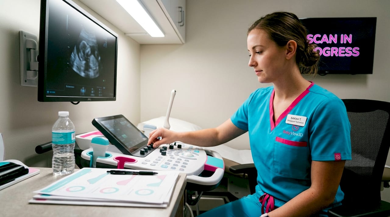
- Ask your sonographer to narrate. Request that they explain what you're seeing in real time, including what is a genuine feature versus a shadow or artifact. A good technician welcomes this conversation and it dramatically increases your understanding.
- Accept that baby's position is outside your control. If the baby's face isn't visible, don't treat it as a failed session. Many studios offer a complimentary return visit if imaging clarity isn't achieved. Ask about this policy before booking.
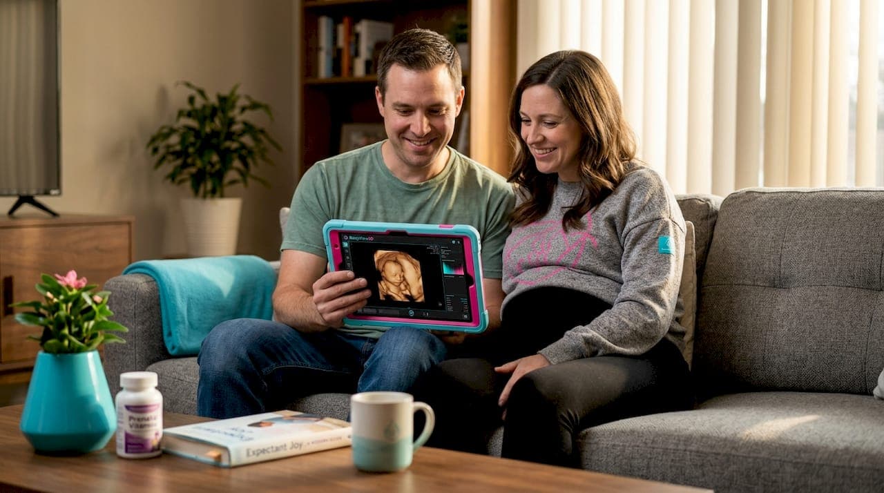
- Bring people who matter. The presence of a partner, parent, or close friend amplifies the emotional experience. A shared moment of seeing the baby's face together is often more memorable than the image itself.
- Keep your clinical scans separate. Your medical anatomy scan is designed to check fetal health and structure. Your visual bonding session is designed to be meaningful and enjoyable. Mixing their purposes in your mind creates confusion about what each one is supposed to tell you.
Pro Tip: After your session, see example images from real clients to understand what various imaging conditions look like. Knowing what a great image versus a limited one looks like helps you process your own session experience.
The emotional power of seeing your baby's face, watching them move, and hearing their heartbeat is genuinely significant. Technology simply gives that experience a visual anchor. The bonding itself is already happening through every kick, every craving, every conversation you have while your hand rests on your belly.
The real value of visual ultrasounds: A balanced perspective
With these practical steps in mind, let's take a step back and offer a candid view of what these scans really add to your journey as expectant parents.
After working with families through thousands of prenatal imaging sessions and seeing their reactions up close, one pattern stands out clearly: the most meaningful moments rarely come from the sharpest image. They come from the instant a parent says, "She has my nose," or when a father sees his baby yawn for the first time and laughs out loud in surprise.
That moment is real. It matters. But it is created by human recognition and emotional connection, not by resolution or technology. Over the past 15-plus years in this field, we've seen parents walk away disappointed because the image "wasn't clear enough" and others leave in tears of joy over a session where the baby barely cooperated. The difference almost always traces back to expectations and to the quality of the human experience in the room.
Here's what we genuinely believe: the biggest risk with visual ultrasounds isn't technical. It's the pressure to have a perfect experience. When parents feel they need a stunning image to prove the bonding is happening, they miss what's already unfolding. The pregnancy itself, the kicks, the growth, the anticipation, that is the bond.
Visual ultrasounds are a beautiful, joyful window into your baby's world. They are worth experiencing. But they are one chapter in a much longer story, not the proof that the story is going well. The unexpected realities of prenatal imaging often include the discovery that what parents treasure most from their session isn't the clearest frame. It's the moment they saw their baby's personality for the first time and simply fell a little more in love.
Go in curious, not demanding. Let the experience be what it is. And trust that the connection you're building with your baby doesn't require perfect lighting.
Ready for your visual ultrasound journey?
If this guide has helped you think more clearly about what visual ultrasounds offer and what to expect, the next step is seeing what a real session can look like for your family.
At BabyView3D, we've spent over 15 years creating safe, meaningful, and genuinely memorable prenatal experiences for families across the United States. Our certified sonographers use advanced HD Live 3D and 4D technology to give you the clearest possible view of your baby while keeping the experience warm, personal, and grounded in professionalism. You can explore our services to find the right package for your stage of pregnancy, see our gallery of real client images to set realistic and exciting expectations, and even browse keepsake products to turn your session into something you'll treasure long after your baby arrives. Your first appointment includes a special introductory offer. Come ready to be amazed.
Frequently asked questions
Are 3D or 4D ultrasounds safe for my baby?
Yes, when performed by trained professionals, 3D and 4D ultrasounds are not associated with known fetal risk. Prudent use by certified sonographers keeps the experience both safe and beneficial.
What if my baby's face or features aren't visible during the session?
This is completely normal. Image clarity depends on fetal position and technical factors, so visibility isn't always guaranteed. Many studios will schedule a follow-up if the initial session doesn't produce clear images.
Is there proof that 3D/4D imaging improves bonding with my baby?
Evidence for consistently improved bonding is limited and results are mixed. Emotional benefits are real but highly individual and context-dependent rather than guaranteed by the technology alone.
Should I schedule visual ultrasounds just for fun?
Non-medical visual ultrasounds are considered investigational and not medically necessary. Always discuss your plan with your healthcare provider so your clinical and bonding scans complement each other appropriately.