Figuring out which ultrasound scans actually matter during pregnancy can feel like trying to read a medical textbook while juggling a million other preparations. Between standard medical scans and the growing world of enhanced 3D and 4D imaging, many expectant parents aren't sure what each milestone involves, what they'll see, or how to make the most of every appointment. Each scan carries both clinical importance and a deeply personal emotional dimension. This guide walks you through every key ultrasound milestone, explains what to expect at each step, and helps you understand how advanced imaging options can turn a medical appointment into a memory you'll treasure for life.
Table of Contents
- Key ultrasound milestones throughout pregnancy
- What to expect: Preparation, procedure, and parent experience
- 2D vs 3D vs 4D ultrasound: Comparing your imaging options
- Safety, prudent use, and current guidance
- A modern perspective: Balancing medical milestones with meaningful memories
- Enhance your ultrasound journey with BabyView3D
- Frequently asked questions
Key Takeaways
| Point | Details |
|---|---|
| Essential ultrasound milestones | Know the timing and purpose of each medically indicated prenatal ultrasound scan. |
| Enhanced imaging options | 3D and 4D ultrasounds provide memorable experiences but are not required for most pregnancies. |
| Preparation matters | Arriving prepared and understanding the process makes each ultrasound smoother and more meaningful. |
| Safety and standards | Ultrasound is considered safe when performed prudently by qualified professionals and within guidelines. |
Key ultrasound milestones throughout pregnancy
Now that you know the value of understanding ultrasound milestones, let's walk through each key scan you'll encounter during pregnancy.
Pregnancy ultrasounds are not one-size-fits-all appointments. Each scan is timed carefully, serves a specific purpose, and offers unique information about your baby's development.
1. First trimester scan (6–10 weeks)
This is your earliest window into your baby's world. The primary goals are confirming viability, establishing gestational age, identifying the number of embryos, and checking the location of the pregnancy. Your provider will look for a heartbeat and measure the embryo to determine your due date more accurately. First trimester ultrasound is the first opportunity to consistently assess anatomy near the end of the trimester, giving your medical team an early baseline to track everything that follows.
2. Nuchal translucency scan (11–14 weeks)
This optional but widely recommended scan measures fluid at the back of the baby's neck. A thicker measurement can indicate an elevated risk for chromosomal conditions like Down syndrome. It's typically combined with blood tests as part of a broader screening program. Not every provider routinely offers this, so ask yours specifically if it's included in your prenatal care plan.
3. Mid-trimester anatomy scan (18–20 weeks)
This is the big one most parents circle on the calendar. The anatomy scan timing is typically 18 to 20 weeks, with potential follow-ups scheduled when additional views are needed. During this scan, your sonographer methodically checks the brain, heart, spine, kidneys, limbs, face, and placenta location. It usually takes between 30 and 60 minutes. This is also when many parents find out their baby's sex, if they choose to.
"The standard mid-trimester anatomy scan typically occurs at 18–20 weeks but may be adjusted depending on individual circumstances."
4. Third trimester scans (28–40 weeks)
Not every pregnancy requires additional ultrasounds after the anatomy scan, but many do. Common reasons include monitoring fetal growth, checking placenta position if placenta previa was noted earlier, assessing amniotic fluid levels, or confirming a baby's position as the due date approaches. Some higher-risk pregnancies may involve weekly scans in the final stretch.
Here is a quick reference for the core milestones:
| Milestone | Timing | Primary purpose |
|---|---|---|
| First trimester scan | 6–10 weeks | Viability, dating, heartbeat confirmation |
| Nuchal translucency | 11–14 weeks | Chromosomal risk screening |
| Anatomy scan | 18–20 weeks | Full structural assessment |
| Growth scan | 28–36 weeks | Size, position, fluid levels |
| Late pregnancy scan | 36–40 weeks | Position check, birth preparation |
Understanding what 4D ultrasound does beyond standard imaging becomes much clearer once you have a firm grasp of where these medical milestones fit.
What to expect: Preparation, procedure, and parent experience
With a clear idea of the major milestones, let's go behind the scenes on what the ultrasound visit itself involves and how you can prepare.
Understanding the mechanics of a scan removes a lot of the mystery and anxiety. Fetal ultrasound uses sound waves to create images and involves little discomfort, typically requiring a full bladder early in pregnancy for better image clarity. For later scans, your bladder state has much less impact on image quality because the baby and amniotic fluid provide enough of a viewing window.
Here's what typically happens during your visit:
- Check-in and preparation: You'll fill out any necessary forms, confirm your gestational age, and be directed to a private room.
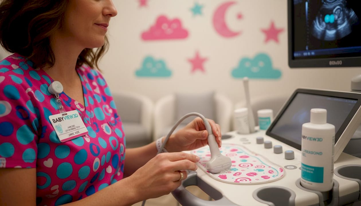
- Gel application: A clear, warm gel is applied to your abdomen. This eliminates air between your skin and the transducer, which improves sound wave transmission.
- Transducer movement: Your sonographer glides the handheld transducer across your belly in various angles to capture different views of the baby and surrounding structures.
- Image capture and measurement: The system records still images and video clips, and the sonographer takes specific measurements based on the scan's purpose.
- Results communication: Depending on the setting, a radiologist or maternal-fetal medicine specialist reviews the images and communicates findings to your OB or midwife, often within a few days.
One thing many parents don't expect is the time spent getting a clear view. Babies move, turn away, or hold their hands in front of their faces. Patience is genuinely part of the process.
Pro Tip: Wear comfortable, two-piece clothing to your ultrasound appointments. Easy access to your abdomen saves time and makes the session more comfortable for everyone.
2D vs 3D vs 4D ultrasound: Comparing your imaging options
Once you know the basics, the natural next question is how enhanced imaging options like 3D and 4D compare with standard ultrasounds.
Standard 2D ultrasound has been the medical backbone of prenatal imaging for decades. It shows cross-sectional slices of your baby in grayscale, which is ideal for clinical measurements and identifying structural features. However, interpreting those flat images takes trained eyes, and most parents find them harder to emotionally connect with compared to more realistic views.
3D and 4D ultrasound data use the same acoustic exposure as standard 2D but display information differently, with 4D capturing movement in real time. That means you're not exposed to more sound energy; the technology simply processes the data into a volumetric image.
Here's how the three main types compare:
| Feature | 2D ultrasound | 3D ultrasound | 4D ultrasound |
|---|---|---|---|
| Image type | Flat, cross-sectional | Still volumetric image | Real-time moving volume |
| Medical use | Primary diagnostic tool | Supplemental anatomy views | Behavioral assessment |
| Emotional impact | Lower for parents | High (first face views) | Very high (movement, expressions) |
| Acoustic exposure | Standard | Same as 2D | Same as 2D |
| Typical availability | All clinical settings | Specialized centers | Specialized centers |
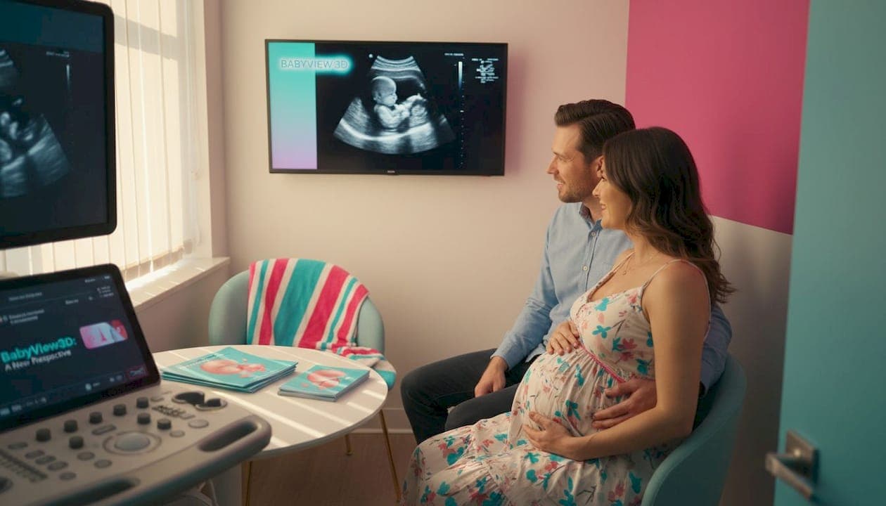
The emotional benefits of 3D and 4D imaging are real. Seeing your baby yawn, stretch, or open and close their mouth during a 4D session creates a level of connection that flat images simply cannot match. For many families, this becomes the moment pregnancy truly becomes tangible.
That said, it's worth being clear: 3D and 4D ultrasounds are primarily experience enhancements. They are not replacements for medically indicated scans. Your anatomy scan will always be the clinical gold standard for structural assessment.
Key benefits parents report from enhanced imaging:
- Stronger emotional bonding before birth
- Sharing the experience with extended family who cannot attend clinical appointments
- Personalized keepsakes including printed images and video clips
- Clearer visualization of familiar facial features
Check out this 3D vs 4D ultrasound guide if you want a deeper side-by-side breakdown, or browse ultrasound package options to understand what different sessions include.
Pro Tip: Ask your provider if they offer 3D or 4D imaging as part of your regular scan, or as a separate add-on. Some practices bundle it in; others refer you to a specialized imaging center.
Safety, prudent use, and current guidance
With so many imaging choices, the topic of safety and expert recommendations deserves a closer look.
The track record of diagnostic ultrasound is genuinely reassuring. No confirmed adverse effects have been reported from present diagnostic ultrasound instruments when used by qualified professionals, though increased intensity or duration may theoretically increase risk. That caveat is why professional guidelines emphasize something called the ALARA principle: as low as reasonably achievable. It means using the minimum exposure needed to get the diagnostic information required.
Where things get nuanced is the world of purely elective, non-medical imaging. Professional policies describe 3D and 4D ultrasound for non-medical keepsakes as not medically necessary and potentially discouraged without clinical context. The U.S. Food and Drug Administration has similarly cautioned against keepsake ultrasound facilities that operate without medical oversight, citing concerns about prolonged exposure without clinical justification.
This does not mean elective imaging is harmful when done responsibly. It means the environment and provider matter enormously.
When evaluating any ultrasound provider, here's what to verify:
- Licensed sonographers with verified credentials and prenatal imaging experience
- Commitment to ALARA principles to minimize exposure duration
- Clear distinction between medical and elective imaging purposes
- No use of Doppler modes for purely entertainment purposes, as these involve higher intensity output
- Transparent communication about what the session includes and excludes
When clinical expertise guides the session, ultrasound can be both a diagnostic tool and a moment of wonder.
Learning how to book a 4D ultrasound responsibly starts with knowing what questions to ask before you ever walk in the door.
A modern perspective: Balancing medical milestones with meaningful memories
Now that you've heard what the experts advise, here's some wisdom from behind the scenes about balancing information with personal experience.
Over more than 15 years of working with expectant families, one pattern stands out clearly: parents who understand the medical purpose of each scan also enjoy the experience of enhanced imaging far more. When you know that your anatomy scan already checked the structural boxes, you can approach a 3D or 4D session with pure wonder rather than anxiety.
Here's the part that surprises many parents: the emotional impact of seeing your baby move in real time is not trivial. Research in prenatal bonding consistently shows that parents who connect visually with their baby before birth report lower anxiety levels and stronger early bonding after delivery. That emotional preparation matters.
But there's a harder truth worth acknowledging. Not every facility offering elective 3D or 4D imaging operates with the same standards. Some prioritize the experience at the expense of clinical rigor. The parents who get the most value are the ones who choose providers that hold both goals equally. They don't treat medical necessity and emotional experience as opposites; they treat them as two parts of the same journey.
Understanding what 4D ultrasound does for families in terms of bonding and preparation goes well beyond a pretty picture. When guided by qualified professionals who respect both the science and the emotion, ultrasound milestones become some of the most memorable moments of your entire pregnancy.
The most enduring takeaway from years in this field: a parent who walks out of an ultrasound session holding a detailed image of their baby's face, knowing everything was reviewed by a certified professional, carries something irreplaceable. That combination of clinical confidence and personal connection is exactly what makes this technology worth understanding deeply.
Enhance your ultrasound journey with BabyView3D
Ready to make the most of your ultrasound milestones? Explore trusted 3D and 4D imaging options designed for your peace of mind.
At BabyView3D, we combine over 15 years of experience with industry-leading HD Live and 8K imaging technology to give you the clearest, most detailed views of your baby before birth. Every session is conducted by certified sonographers who prioritize both clinical standards and the emotional experience your family deserves.
Whether you're approaching your anatomy scan or planning a dedicated bonding session in your third trimester, our team is here to guide you every step of the way. View BabyView3D services to explore everything we offer, or shop BabyView3D packages to find the session that fits your timeline and wishes. First-time visitors can also access a limited-time discount on their initial appointment.
Frequently asked questions
When is the best time for a 3D or 4D ultrasound during pregnancy?
Most providers recommend scheduling 3D or 4D ultrasounds between 26 and 32 weeks, when the baby has developed enough fat beneath the skin to show clear facial features while still having enough room to move within the womb.
Is ultrasound safe for my baby and me?
Diagnostic ultrasounds used by qualified professionals have not shown harmful effects for mother or fetus. No confirmed adverse effects have been reported from diagnostic ultrasound instruments when operated by trained professionals following established guidelines.
Do I need a full bladder for every ultrasound?
Early ultrasounds may require a full bladder for better imaging clarity, but later pregnancy scans generally do not. Early pregnancy prep may include drinking water for a more visible uterus, while later scans rely on amniotic fluid for a clear viewing window.
Are 3D and 4D ultrasounds medically necessary?
3D and 4D ultrasounds are usually considered non-essential and primarily for experience, unless your provider identifies a specific clinical reason. 3D and 4D for non-medical purposes is considered not medically necessary by professional policies, though many families choose them as a meaningful addition to their prenatal journey.