4D ultrasound is defined as real-time, live-video 3D imaging of the womb, and it surpasses 3D ultrasound for fetal movement because it captures continuous motion rather than a frozen still. Where a 3D scan delivers a single detailed snapshot, 4D adds the dimension of time, letting you watch your baby kick, yawn, blink, and stretch as it happens. This distinction matters both emotionally and clinically. Parents gain a genuine first look at their child's personality, and clinicians gain behavioral data that static images simply cannot provide. Understanding why 4D imaging leads in fetal movement visualization helps you make a more informed choice before booking your next prenatal scan.
Why 4D surpasses 3D for fetal movement: the core difference
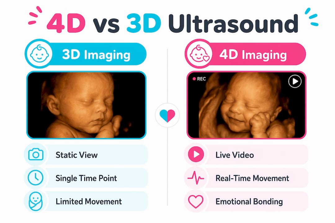
The fundamental reason 4D imaging outperforms 3D for movement comes down to one word: time. A 3D scan stitches sound waves into a single three-dimensional still image. That image can show remarkable facial detail, but the moment your baby moves, the picture is already outdated.
4D adds the fourth dimension, time, by rendering those same sound waves into a continuous stream of frames. The result is live video. You see your baby's arm sweep across its face, its chest rise with a practice breath, and its mouth open in a yawn, all in real time.
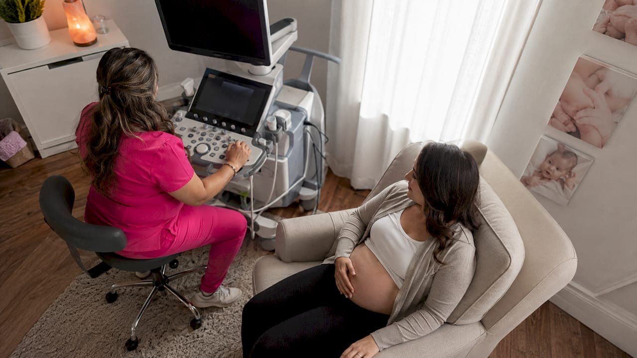
Here is how the process works step by step:
- Sound wave emission: The transducer sends high-frequency sound waves into the uterus.
- Echo capture: Returning echoes are collected and converted into raw data.
- 3D rendering: Software assembles the data into a three-dimensional surface image.
- Time layer added: In 4D mode, the software repeats this rendering process many times per second, producing a live video feed.
- Display output: The video streams directly to the monitor so you can watch movement as it occurs.
The underlying ultrasound exposure is identical for 3D and 4D scans. The difference is entirely in how the software processes and displays the data, not in any additional energy directed at the baby.
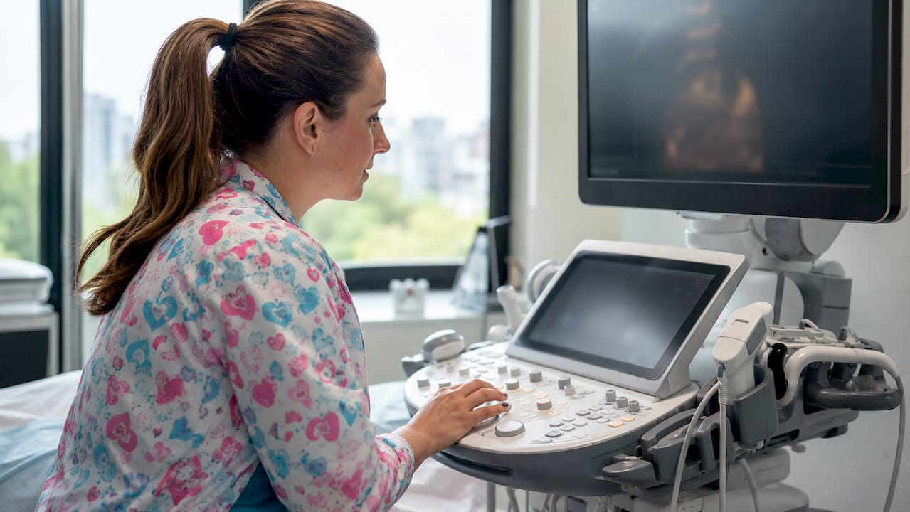
Pro Tip: The sonographer's skill has a larger impact on image quality than the equipment brand. Ask your provider how many 4D sessions they perform each week before you book.
What are the real benefits of 4D ultrasound for parents?
The advantages of 4D imaging for fetal movement visualization fall into two clear categories: emotional and clinical. Both are significant, and neither is available through a static 3D image.
Emotional connection before birth
Real-time fetal movement reduces parental anxiety and builds emotional attachment in a way that still images cannot replicate. Watching your baby suck its thumb or respond to sound creates a sense of relationship before delivery. This is not a minor benefit. Research on prenatal emotional bonding consistently links early parental attachment to better postpartum outcomes for both parent and child.
4D imaging also lets the whole family participate. Partners, siblings, and grandparents can watch the baby move in real time on a monitor, making the experience shared rather than solitary.
Clinical insights that static images miss
Clinicians find 4D ultrasound valuable for functional behavioral assessment including swallowing patterns, limb activity, and breathing movements. These behaviors cannot be assessed from a single frozen frame. A 3D still might confirm a structural feature, but it tells you nothing about whether the baby is practicing swallowing or moving its limbs symmetrically.
The numbered list below summarizes the six core advantages of 4D over 3D for fetal movement:
- Live kicking and stretching: You observe actual movement sequences, not a single frozen position.
- Facial expression capture: Yawning, blinking, and smiling are visible in motion, not just implied by structure.
- Breathing pattern observation: Practice breathing movements confirm lung activity in real time.
- Swallowing behavior: Clinicians can assess swallowing function, a key developmental marker.
- Anomaly detection support: Asymmetric limb movement or absent reflexes become visible during live observation.
- Parental reassurance: Watching a baby move actively provides immediate, tangible confirmation of wellbeing.
"Seeing real-time interaction reduces anxiety and helps parents connect emotionally before birth." — Ashwin Scans, Benefits of 4D Scans for Parents
A 3D/4D scan also monitors growth, placenta position, and blood flow as part of comprehensive prenatal care. The 4D component adds a behavioral layer on top of that structural data.
When is the best time for a 4D ultrasound vs. 3D?
Timing matters more for 4D than for 3D because movement visibility depends on the baby's size, position, and the amount of amniotic fluid surrounding it.
| Factor | 3D Ultrasound | 4D Ultrasound |
|---|---|---|
| Primary output | Static 3D image | Live video of movement |
| Optimal gestational window | 24–32 weeks | 26–30 weeks for clearest movement |
| Session length | 20–30 minutes | 20–30 minutes |
| Typical elective cost | $99–$149 | Often bundled with 3D at same price |
| Movement visibility | Limited to position at capture | Continuous real-time observation |
| Clinical use | Structural assessment | Structural plus behavioral assessment |
The optimal imaging window is 26–30 weeks for the clearest fetal movement and facial feature views. Before 24 weeks, the baby lacks the fat deposits that define facial features. After 32 weeks, limited space in the uterus reduces both movement range and image clarity.
Key session factors that affect 4D quality include:
- Amniotic fluid level: More fluid around the baby's face produces sharper images.
- Fetal position: A baby facing the uterine wall will block the view regardless of technology used.
- Maternal body composition: Adipose tissue can reduce sound wave penetration and image sharpness.
- Hydration: Drinking water in the 24 hours before your session can improve fluid levels and image quality.
Elective 3D/4D sessions typically cost $99–$149, and 4D video capability is almost always included as a software feature rather than a separate hardware upgrade. That means you rarely pay more for the live video component.
What are the limitations of 4D ultrasound?
4D imaging is not without constraints. Understanding them helps you set realistic expectations and get the most from your session.
- Baby's position is the biggest variable. If the baby faces away or has a limb in front of its face, no amount of technology will produce a clear frontal view. The sonographer may ask you to walk around or return for a second look.
- Late pregnancy reduces clarity. Beyond 32 weeks, reduced amniotic fluid and limited space make fetal movement harder to capture clearly.
- Low amniotic fluid limits imaging. Oligohydramnios, a condition of reduced amniotic fluid, significantly reduces 4D image quality regardless of operator skill.
- Operator skill determines results. The quality of results depends primarily on the sonographer's experience and the baby's cooperation, not on the software label used.
- Marketing terms add confusion. Labels like 5D or HD Live describe visual rendering enhancements layered on top of standard 4D video. They do not represent a different physical imaging technique or provide additional diagnostic capability.
- 4D is not medically required. Elective 4D sessions complement your standard prenatal care. They do not replace the diagnostic 2D scans your OB or midwife orders. Think of 4D as an addition to your prenatal experience, not a substitute for clinical monitoring.
Pro Tip: Book your elective 4D session between weeks 26 and 28 for the best balance of image clarity, fetal size, and movement activity. Drink at least 64 ounces of water daily in the two days before your appointment.
Understanding ultrasound resolution improvements can also help you evaluate what to expect from different imaging formats before you arrive.
Key Takeaways
4D ultrasound surpasses 3D for fetal movement because it adds the dimension of time, converting static images into live video that captures kicking, yawning, and breathing in real time.
| Point | Details |
|---|---|
| Core 4D advantage | 4D adds real-time video to 3D imaging, showing live fetal movement instead of a frozen still. |
| Best timing | Schedule between 26–30 weeks gestation for the clearest movement and facial feature views. |
| Safety is equal | 4D uses identical sound wave exposure as 3D and 2D scans; the difference is software rendering only. |
| Clinical value | 4D enables observation of swallowing, breathing, and limb activity that static 3D images cannot assess. |
| Manage expectations | Baby position and amniotic fluid level affect image quality more than the technology label used. |
What I've learned from watching families see their baby move for the first time
I have been in the room when parents see their baby yawn on a 4D monitor. The reaction is never subtle. There is a specific moment, usually around the 90-second mark of a session, when the parent stops watching the screen and starts talking to the baby. That shift from observer to parent is something a still image rarely triggers.
What I find underappreciated is how 4D imaging changes the father's experience specifically. Partners who feel disconnected during pregnancy because they cannot feel movement often describe a 4D session as the moment the pregnancy became real for them. That is not a small thing.
My practical advice: do not wait until 32 weeks hoping for better image quality. The baby is often too large and too settled by then. The 26–28 week window is genuinely the sweet spot, and I have seen far too many families miss it chasing a later date.
One more thing worth saying clearly: 4D does not replace your standard prenatal care. Your OB's 2D anatomy scan at 20 weeks is doing diagnostic work that 4D is not designed to replicate. Use 4D for what it does best, which is giving you a live, moving introduction to your child before birth.
— LENIER
See your baby move with Bbview3d
Bbview3d has spent more than 15 years helping families across the United States experience their baby's first movements in stunning detail. Every session combines 3D and 4D imaging with HD Live technology, delivered by certified sonographers in a comfortable, family-centered environment.
Sessions are available at multiple locations, and first-appointment pricing makes it easy to book without hesitation. Whether you want to capture your baby's yawn, share the moment with your whole family, or simply see your child move for the first time, Bbview3d's prenatal ultrasound services are built for exactly that. Book your session today and bring the whole family.
FAQ
What is the main difference between 3D and 4D ultrasound?
3D ultrasound produces a static three-dimensional image, while 4D ultrasound adds the dimension of time to create live video. The result is that 4D shows fetal movement in real time, including kicking, yawning, and facial expressions.
Is 4D ultrasound safe for my baby?
Yes. 4D imaging uses the same sound wave technology as 3D and 2D ultrasounds. The difference between formats is in software rendering, not in the level of energy directed at the baby.
When should I schedule a 4D ultrasound?
The optimal window is 26–30 weeks gestation. This period offers the best balance of fetal size, amniotic fluid volume, and movement activity for clear imaging.
How much does a 4D ultrasound session cost?
Elective sessions typically run $99–$149, and 4D video is usually included in the same package as 3D imaging without an additional hardware cost.
Does 4D ultrasound replace my standard prenatal scans?
No. 4D elective sessions complement your OB-ordered 2D anatomy scans but do not replace them. Standard 2D scans perform diagnostic assessments that 4D is not designed to replicate.