Pregnancy comes with a flood of appointments, acronyms, and scan types that can leave any expectant parent feeling genuinely lost. When your doctor mentions high-detail fetal imaging, you might picture a standard ultrasound with a slightly sharper screen. The reality is far more specific than that. High-detail fetal imaging refers to a category of advanced prenatal scanning techniques, including high-resolution ultrasound, 3D and 4D imaging, fetal MRI, and fetal echocardiography, each designed to reveal structural and developmental information that routine scans simply cannot capture. This guide breaks down what these methods involve, when they happen, and why they matter for your baby's health and your peace of mind.
Table of Contents
- Key takeaways
- What high-detail fetal imaging actually means
- When and why high-detail scans are performed
- Benefits of high-detail fetal imaging
- The latest advances in fetal imaging technology
- My honest take on what this means for families
- See your baby like never before with Bbview3d
- FAQ
Key takeaways
| Point | Details |
|---|---|
| More than a clearer picture | High-detail fetal imaging is a category of specialized techniques with distinct clinical and bonding purposes. |
| Timing varies by method | Most detailed anatomy scans occur between 18 and 22 weeks, with select first-trimester scans for high-risk pregnancies. |
| Multiple technologies exist | Methods range from 3D/4D ultrasound to fetal MRI and fetal echocardiography, each revealing different structures. |
| AI is reshaping the field | Automated measurement tools now reduce human variability and improve early detection of growth issues. |
| Image quality has real limits | Fetal position, maternal factors, and timing all affect what any scan can show, regardless of the technology used. |
What high-detail fetal imaging actually means
The standard term clinicians use is "advanced fetal ultrasound" or "level II sonography," though the broader category includes several distinct technologies. Understanding each one helps you ask better questions at your next appointment.
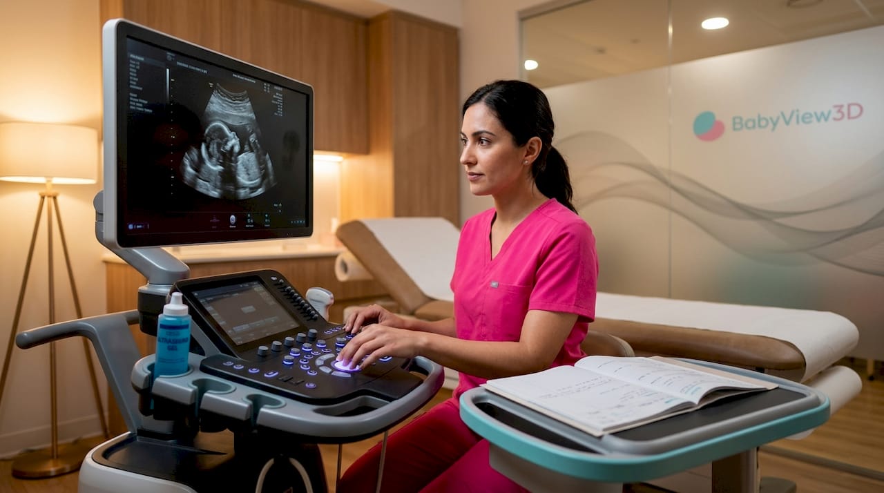
High-resolution ultrasound is the foundation. Unlike routine first-trimester dating scans, a level II anatomy survey examines structures like the four-chamber heart view, brain anatomy, abdominal organs, and spine in systematic detail. It is noninvasive, uses sound waves, and carries no radiation risk, making it the workhorse of prenatal assessment.
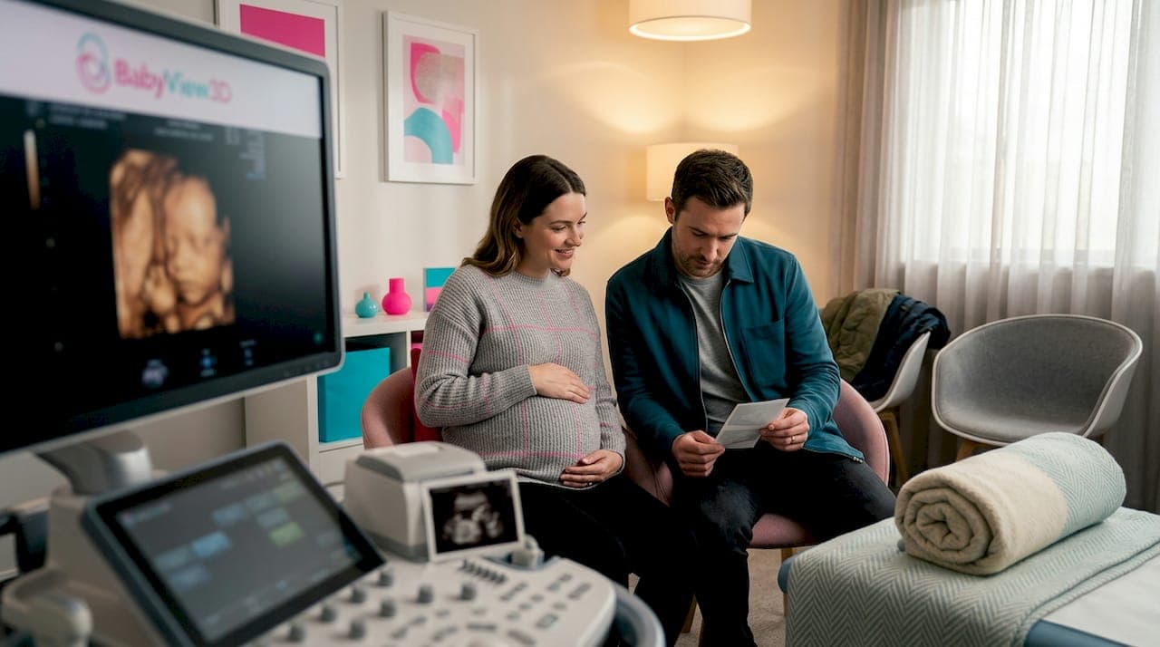
3D and 4D ultrasound take the same sound wave technology and reconstruct it differently. 3D adds real-time movement, turning flat echoes into a rendered surface image. 4D adds the time dimension, so you watch your baby yawn, stretch, or open a hand in motion. Clinicians primarily rely on 2D for diagnostic measurements, but 3D and 4D add meaningful context for both structural review and parental bonding.
Fetal MRI operates on a completely different principle. At high field strengths of 4T and above, fetal MRI achieves spatial resolutions between 10 and 100 micrometers, which means brain structures invisible on standard ultrasound become visible. It is used selectively, most often when ultrasound findings are inconclusive or when brain development requires closer scrutiny.
Fetal echocardiography focuses entirely on the heart. A pediatric cardiologist or specialist sonographer maps blood flow, valve function, and cardiac chamber structure in detail that a routine scan cannot provide. It is typically ordered when a family history of heart defects, maternal diabetes, or an unusual four-chamber view raises concern.
Here is a summary of the key techniques and their primary uses:
- High-resolution (level II) ultrasound: systematic scan of all major fetal organ systems, typically at 18 to 22 weeks
- 3D/4D ultrasound: surface rendering for structural review and parental bonding experiences
- Fetal MRI: detailed brain and soft-tissue imaging when ultrasound findings need clarification
- Fetal echocardiography: dedicated cardiac assessment for suspected heart anomalies
- AI-assisted imaging: automated measurement and anomaly detection layered onto existing modalities
Pro Tip: If your provider mentions a level II scan, ask specifically whether it includes a 4-chamber cardiac view. That single view is one of the most important structural checkpoints in the entire anatomy survey.
When and why high-detail scans are performed
The timing of a specialized prenatal scan is not arbitrary. Each window in pregnancy offers different structural visibility, and the reason for the scan shapes which week makes sense.
-
First trimester (12 to 14 weeks): Indication-driven anatomy scans at this stage are reserved for high-risk situations, including advanced maternal age, a prior pregnancy with chromosomal anomalies, or abnormal serum screening results. These early scans can detect certain structural issues but are not a substitute for second-trimester surveys.
-
Second trimester (18 to 22 weeks): This is the primary window for detailed anatomy imaging. The fetus is large enough for reliable organ measurement, yet the amniotic fluid volume and fetal positioning generally allow clear access. Most level II scans, fetal echocardiograms, and 3D sessions are scheduled here.
-
Third trimester: Additional detailed scans may be ordered to monitor growth in pregnancies complicated by gestational diabetes, hypertension, or previously identified anomalies. Fetal position and reduced fluid volume can make imaging more challenging at this stage.
The reasons a provider orders advanced imaging also vary widely. Routine screening differences between populations, maternal factors like obesity or uterine fibroids, and suspected findings on a standard scan all serve as triggers. Fetal position and maternal body factors are among the most significant variables affecting what any given scan can show. Even the most advanced technology cannot override physics.
"Advanced fetal imaging is most powerful when paired with clinical context. A finding on a scan means something different depending on the full picture of that pregnancy."
Some parents also choose high-detail prenatal scans for elective reasons, particularly 3D and 4D sessions that prioritize the bonding experience. These are distinct from medically ordered surveys in their purpose, though they use the same underlying technology. Understanding this difference helps you set the right expectations before you walk in the door.
Benefits of high-detail fetal imaging
The practical value of advanced prenatal imaging falls into two broad categories: medical and emotional. Both are legitimate, and neither one cancels out the other.
On the medical side, the benefits are concrete:
- Structural anomaly detection: Detailed scans can identify issues with the brain, heart, kidneys, spine, and limbs that routine screens miss entirely. Earlier detection creates more preparation time for families and care teams.
- Delivery planning: When an anomaly like a lung malformation is identified, imaging guides delivery planning to put the right specialists in place at the right hospital before the baby arrives.
- Tailored prenatal care: Findings from detailed scans directly inform decisions about frequency of monitoring, medication adjustments, and activity restrictions through the rest of pregnancy.
- Fetal heart assessment: A fetal echocardiogram can diagnose complex congenital heart conditions before birth, which dramatically changes how the delivery team prepares.
On the emotional side, the impact is equally real. Seeing your baby's face, hands, or a recognizable expression through 3D and 4D imaging creates a connection that flat 2D scans rarely achieve. For parents navigating anxiety or a complicated pregnancy, seeing a detailed, recognizable image can be genuinely stabilizing.
Pro Tip: Drink water consistently in the 24 to 48 hours before a 3D or 4D session. Hydration improves amniotic fluid clarity, which directly affects surface image quality.
One important limitation deserves honest acknowledgment. High-detail does not mean perfect. Image quality depends heavily on fetal position, how far along you are, and your individual body. Technology sets the ceiling, but real-world conditions determine where you land below it.
The latest advances in fetal imaging technology
2026 has brought meaningful progress to the field, and knowing what is available helps you have better conversations with your provider.
AI-assisted measurement is now integrated into many clinical ultrasound platforms. Automated biometry systems achieve measurement precision comparable to experienced sonographers, which matters most in settings where operator experience varies. These tools can flag growth concerns earlier and reduce the chance that a subtle finding gets missed.
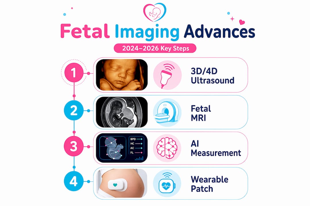
Beyond measurement, deep learning models trained on fetal brain and craniofacial structures are improving the automated detection of abnormal anatomy. These systems do not replace clinical judgment. They add a layer of consistency that benefits patients, particularly in high-volume imaging centers.
| Technology | What it adds | Current status |
|---|---|---|
| AI biometry | Automated measurements, growth flagging | Clinically available |
| High-field fetal MRI | Brain microstructure detail at 10-100 micrometers | Specialized centers |
| Wearable ultrasound patch | Continuous fetal monitoring over hours or days | Emerging, high-risk use |
| HD Live 3D/4D ultrasound | Realistic surface rendering for bonding and review | Widely available |
Wearable ultrasound patches represent one of the more interesting developments for high-risk pregnancies. Continuous fetal monitoring over extended periods captures developmental trends that snapshot scans simply cannot. Still, these patches supplement standard scans rather than replace them. The periodic detailed anatomy survey remains the benchmark. For more on where these technologies are heading, the ultrasound trends in 2026 piece covers the clinical and consumer shifts in real depth.
Questions worth asking your provider: Does your imaging center use AI-assisted measurement tools? If a finding on my anatomy scan is unclear, is fetal MRI available as a next step? For high-risk pregnancies, would continuous monitoring be appropriate in my case?
My honest take on what this means for families
I've spent enough time watching parents walk into imaging appointments with unrealistic expectations on both ends of the spectrum. Some expect a cinema-quality reveal. Others are braced for disaster. Neither posture actually serves you.
What I've found is that high-detail fetal imaging is most valuable when you understand what each method is designed to do before you walk in. A level II anatomy scan is not a keepsake session. A 3D bonding scan is not a substitute for a medical survey. Knowing the difference means you show up to each appointment with the right questions and the right frame of mind.
The advances in AI and fetal MRI are genuinely impressive. But the part that matters most for most families is still the skill and attentiveness of the person holding the transducer. Technology raises the floor. Caring, experienced sonographers raise the ceiling.
My encouragement: be present at these appointments, ask about what you are seeing in real time, and follow up on anything that was not fully explained. The image on the screen is your child. You are allowed to ask questions.
— LENIER
See your baby like never before with Bbview3d
Bbview3d has spent over 15 years helping expectant families experience prenatal imaging the way it should feel: clear, personal, and unforgettable. Using HD Live technology with 3D and 4D ultrasound in resolutions up to 8K, Bbview3d goes far beyond what a standard clinic visit provides. Certified sonographers guide every session, and flexible packages are designed around your timeline and preferences. Whether you are looking for a medical-quality scan experience or a bonding session you will carry with you forever, the prenatal imaging services at Bbview3d are worth exploring. First-time visitors can also browse the session gallery to see the image quality for themselves before booking.
FAQ
What is high-detail fetal imaging in simple terms?
High-detail fetal imaging is an umbrella term for advanced prenatal scanning techniques, including level II ultrasound, 3D/4D imaging, fetal MRI, and fetal echocardiography, that reveal structural and developmental details beyond what routine scans can show.
When is a detailed anatomy scan typically performed?
Most detailed anatomy scans are performed between 18 and 22 weeks of pregnancy, when the fetus is large enough for reliable structural assessment. First-trimester versions between 12 and 14 weeks are reserved for high-risk cases.
What does fetal echocardiography check for?
Fetal echocardiography is a specialized scan focused exclusively on the baby's heart. It assesses chamber structure, valve function, and blood flow patterns, and is typically ordered when a family history of heart conditions or an unclear standard cardiac view is present.
How does AI improve fetal imaging?
AI tools automate biometric measurements and standard-plane detection during scans. AI biometry systems achieve precision comparable to experienced sonographers, which helps reduce variability and supports earlier detection of growth abnormalities.
Can high-detail imaging guarantee clear pictures of my baby?
No. Even the best technology cannot override real-world variables. Image clarity depends on fetal position, amniotic fluid volume, gestational age, and maternal body factors, all of which affect what any scan can realistically show.