

Most parents don't realize that ultrasound serves a far bigger role than confirming a heartbeat. Understanding why families choose ultrasound means looking at the full picture: clinical safety checks, emotional reassurance, and the irreplaceable moment of seeing your baby's face before birth. This guide covers every reason expectant parents schedule ultrasounds, from the medically necessary anatomy scan to the elective session that lets grandparents see the baby's features for the first time, so you can make informed, confident decisions throughout your pregnancy.
Table of Contents
- Key takeaways
- Why families choose ultrasound during pregnancy
- The emotional reasons parents value ultrasound
- Understanding ultrasound types and timing
- Misconceptions about in-person ultrasound
- My take on what ultrasound really means for families
- See your baby in stunning detail with Bbview3d
- FAQ
Key takeaways
| Point | Details |
|---|---|
| More than a medical test | Ultrasound provides clinical data, emotional reassurance, and lasting family memories all in one appointment. |
| Timing affects accuracy | Scans performed at the right pregnancy stage deliver more reliable fetal dating and anomaly detection. |
| Emotional bonding is real | Seeing detailed images reduces parental anxiety and strengthens attachment before birth. |
| Not all conditions are detectable | Ultrasound is powerful but limited; some conditions require follow-up testing or develop after the scan. |
| Provider quality matters | Certified sonographers and qualified studios deliver safer, more meaningful ultrasound experiences. |
Why families choose ultrasound during pregnancy
Most families think of ultrasound as a box to check on the prenatal care list. In reality, it does several very different jobs at once, and knowing those jobs helps you prepare for each appointment with the right mindset.
The anatomy scan: what it actually shows
The mid-pregnancy anatomy scan, typically scheduled between 18 and 22 weeks, is the most thorough single view of your baby's development you will receive during pregnancy. Sonographers measure the brain, heart, spine, limbs, kidneys, and face. They also check the placenta's position and the amount of amniotic fluid surrounding your baby.
Here is what this scan evaluates:
- Fetal brain and spine structure, checking for neural tube concerns
- Four-chamber heart view, screening for major cardiac differences
- Limb length and bone development, confirming growth is on track
- Placenta location, identifying placenta previa before delivery planning
- Amniotic fluid levels, which reflect kidney function and swallowing behavior
| What ultrasound can detect | What it may miss |
|---|---|
| Major structural differences in organs | Minor heart defects that develop later |
| Placenta position and cord insertion | Soft tissue conditions without structural change |
| Estimated fetal weight and growth rate | Genetic conditions without physical markers |
| Fetal presentation and position | Conditions that develop after the scan date |
Pro Tip: Drink water before your anatomy scan but avoid a painfully full bladder. A moderately full bladder helps with image quality in the second trimester without the discomfort needed for first-trimester scans.
Beyond the anatomy scan, ultrasound plays a critical supporting role after genetic screening results. When cell-free DNA (cfDNA) screening returns elevated risk results, an anatomic ultrasound survey can detect physical markers that cfDNA may miss, helping your provider determine whether diagnostic testing like amniocentesis is warranted. It also confirms fetal position and refines your due date, since ultrasound timing accuracy drops significantly when scans are performed late in the third trimester rather than during the optimal mid-pregnancy window.
The emotional reasons parents value ultrasound
Clinical necessity alone does not explain why so many families schedule multiple scans or choose elective sessions on top of their standard prenatal care. The emotional weight of a pregnancy ultrasound is real and well-documented.
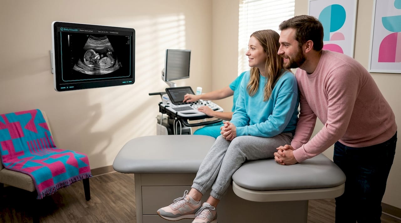
The 20-week scan is often the first time parents see a fully formed baby rather than a flickering shape on a screen. That moment combines joy, awe, and a type of relief that no blood test or report can replicate. For parents who have experienced pregnancy loss or fertility treatment, the reassurance of seeing a healthy heartbeat and a baby that looks like a baby carries enormous psychological weight. Families often choose additional elective ultrasounds for exactly this reason, especially after fertility challenges or a previous miscarriage where anxiety is heightened throughout a subsequent pregnancy.
Here is why the emotional side of ultrasound matters as much as the clinical side:
- It turns an abstract pregnancy into a recognizable baby, accelerating parental attachment before birth
- It gives partners, siblings, and grandparents a shared visual experience that builds family connection
- It creates keepsake images and videos that families return to for years after delivery
- It reduces the uncertainty that drives pregnancy anxiety, replacing "I hope everything is okay" with "I saw the heartbeat"
Pro Tip: If anxiety is high before a scan, ask your provider to narrate what they are seeing in real time. Hearing "here are the four chambers of the heart, and they all look normal" is far more calming than waiting in silence for a summary.
Advanced imaging technologies like 3D, 4D, and HD Live make these emotional benefits even more pronounced. When you can see your baby yawn, stretch, or turn their head, the bonding experience shifts from looking at a medical image to genuinely meeting your child. That distinction matters to families, and it is one of the core reasons why parents prefer in-person ultrasound at a specialized studio over a printed black-and-white image from a clinical appointment. You can learn more about how HD Live imaging transforms the experience if you are considering an elective session.
Understanding ultrasound types and timing
Knowing which scan you are getting and when it happens helps you prepare practically and emotionally. The standard prenatal schedule in the United States typically follows this sequence:
- First trimester dating scan (6 to 9 weeks): Confirms the pregnancy is in the uterus, establishes a heartbeat, and provides an early due date estimate.
- Nuchal translucency scan (11 to 14 weeks): Measures fluid at the back of the baby's neck as part of first-trimester chromosomal screening.
- Mid-pregnancy anatomy scan (18 to 22 weeks): The comprehensive structural survey described above.
- Growth scans (28 weeks onward): Recommended for high-risk pregnancies or when earlier scans raised questions about fetal growth.
- Elective or keepsake ultrasounds: Scheduled by parents at any point, typically between 24 and 32 weeks for the best 3D imaging results.
| Scan type | Purpose | Who typically schedules it |
|---|---|---|
| Dating scan | Confirm viability and due date | OB-GYN or midwife |
| Anatomy scan | Full structural assessment | Standard prenatal care |
| Growth scan | Monitor size and fluid | High-risk pregnancies |
| Elective 3D/4D | Bonding and keepsake imagery | Parents, through a boutique studio |
When timing matters, it really matters. Clinicians recommend scans within specific gestational windows because accuracy drops outside those ranges. A due date calculated from a third-trimester scan, for example, can be off by two to three weeks because fetal growth rates vary more later in pregnancy. Scheduling your anatomy scan before 22 weeks gives the sonographer time to arrange a follow-up appointment if any structures need a second look. For elective 3D sessions, the sweet spot is 26 to 30 weeks. The baby has enough fat under the skin to produce detailed facial features, but still has space in the uterus to shift into a viewable position. Understanding the prenatal ultrasound packages guide can help you decide which sessions fit your pregnancy timeline.
Misconceptions about in-person ultrasound
Several common misunderstandings keep expectant parents from getting the most out of their ultrasound appointments or from making confident decisions about elective sessions.
Misconception 1: Ultrasound uses radiation. It does not. Ultrasound uses sound waves, not ionizing radiation, making it safe for repeated use throughout pregnancy without the risks associated with X-rays or CT scans. This is a fundamental distinction that makes ultrasound the preferred method for repeated fetal assessment.
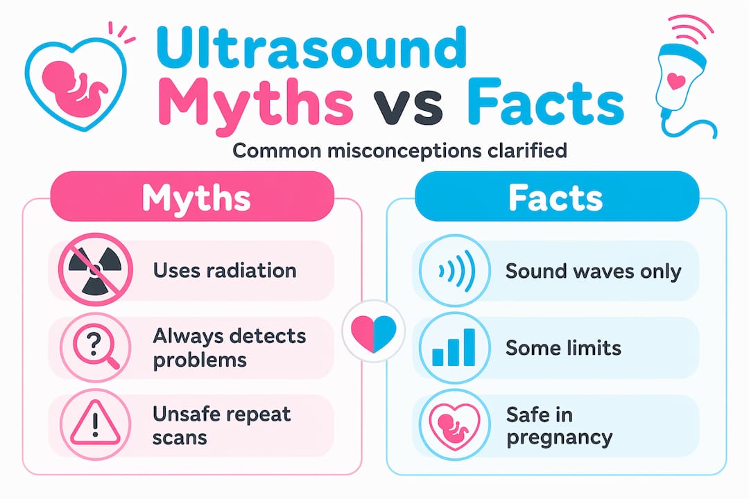
Misconception 2: A normal ultrasound means nothing is wrong. Ultrasound provides critical information, but not all conditions can be detected through imaging alone. Some structural differences are subtle, some develop after the scan date, and some conditions leave no visual marker at all. A reassuring scan is genuinely good news, but it works alongside other tests rather than replacing them.
Misconception 3: Any provider offering ultrasound delivers the same experience. The quality of your ultrasound depends heavily on the skill of the sonographer, the caliber of the equipment, and the protocols followed. Qualified, credentialed providers differentiate diagnostic scans from casual viewing sessions, and that distinction directly affects the accuracy and safety of your results.
"Ultrasound results require context. A finding on a scan is a data point, not a diagnosis. Families who understand this leave appointments better prepared for whatever comes next, whether that is a follow-up scan, a specialist referral, or simply peace of mind."
Choosing a provider who communicates clearly, uses up-to-date equipment, and holds proper certifications protects both the quality of your imaging and the accuracy of your expectations.
My take on what ultrasound really means for families
I've watched hundreds of families walk into an ultrasound session nervous and walk out transformed. That shift is not a small thing. In my experience, the parents who get the most from their ultrasound appointments are the ones who understand that these scans serve two entirely different needs at once: clinical information and emotional confirmation.
What I've learned is that most of the anxiety surrounding prenatal imaging comes from not knowing what to expect. When parents understand that the anatomy scan is a structured measurement survey designed to flag concerns early, the 20-week scan anxiety drops significantly. They walk in informed rather than fearful.
My honest opinion is that elective ultrasound sessions, done at the right time with a qualified provider, are one of the most undervalued parts of the prenatal experience. Not because they replace clinical care, but because they give families something clinical care rarely has time to offer: a slow, unrushed moment to meet their baby. I've seen that moment change the tone of an entire pregnancy. It makes the abstract concrete and the anxious hopeful. That is worth planning for.
— LENIER
See your baby in stunning detail with Bbview3d
If you are ready to experience the emotional side of prenatal ultrasound, Bbview3d offers sessions that go far beyond the standard clinical scan. With over 15 years of experience and certified sonographers using 3D, 4D, and 8K HD Live technology, Bbview3d creates a warm, unhurried environment where families can truly connect with their baby before birth. Whether you want a bonding session after fertility treatment, a keepsake experience to share with the whole family, or simply a longer, more personal look at your growing baby, Bbview3d's ultrasound services are designed with exactly that in mind. Explore packages, see real family testimonials, and book your first appointment at Bbview3d.
FAQ
Why do families choose ultrasound beyond medical need?
Many families choose elective ultrasounds for emotional bonding, keepsake memories, and reassurance, especially after fertility treatment or pregnancy loss. The visual experience of seeing a baby in detail strengthens parental attachment in ways clinical reports cannot replicate.
Is prenatal ultrasound safe for repeated use?
Yes. Ultrasound uses sound waves rather than radiation, making it safe for repeated fetal assessment throughout pregnancy. It is the standard method for monitoring fetal health precisely because of this safety profile.
When is the best time to schedule a 3D elective ultrasound?
The optimal window for 3D and 4D elective ultrasounds is between 26 and 32 weeks. At this stage, the baby has developed enough facial fat for detailed imaging while still having room to move into a clear viewing position.
Can a normal ultrasound guarantee a healthy pregnancy?
No. Ultrasound provides critical data but cannot detect all conditions. Some anomalies are subtle, develop after the scan, or require genetic testing to identify. A reassuring scan is genuinely positive news, but it works best as part of a complete prenatal care plan.
How do I choose a qualified ultrasound provider?
Look for certified sonographers, up-to-date imaging equipment, and transparent communication about what the scan can and cannot show. For elective sessions, choose studios with professional credentials and a clear policy of recommending physician involvement for any clinical concerns.