If you've ever stared at a prenatal scan and wondered why some images look crystal clear while others look like static, the answer comes down to ultrasound resolution. What is ultrasound resolution, exactly? It's not simply about sharpness. Resolution in ultrasound is a combination of three distinct technical properties, each affecting what the sonographer can see, measure, and assess about your baby. Understanding these properties helps you make sense of what you see on screen and what questions to ask at your next prenatal visit.
Table of Contents
- Key takeaways
- What is ultrasound resolution in prenatal imaging
- How frequency shapes what you see
- Super-resolution ultrasound and what it means for prenatal care
- How resolution affects your prenatal ultrasound experience
- My honest take on resolution and what actually matters
- See your baby with exceptional clarity at Bbview3d
- FAQ
Key takeaways
| Point | Details |
|---|---|
| Three types of resolution | Axial, lateral, and temporal resolution each serve a different purpose in prenatal imaging. |
| Frequency involves trade-offs | Higher frequency improves detail but reduces how deep the sound waves can reach. |
| Super-resolution is emerging | New technology can achieve 10-micrometer resolution, far beyond conventional ultrasound limits. |
| Settings matter as much as hardware | Adjusting focal zones and frame rates can noticeably improve image clarity without changing equipment. |
| Resolution shapes your experience | The clarity of 3D and 4D images depends directly on how well all three resolution types are optimized. |
What is ultrasound resolution in prenatal imaging
The word "resolution" gets used loosely, so let's be specific. Ultrasound spatial resolution consists of three measurable properties: axial, lateral, and temporal resolution. Together, they determine how much detail a sonographer can extract from any given scan. Think of them as three separate lenses, each focused on a different dimension of the image.
Axial resolution measures how well the system distinguishes two objects that are positioned one in front of the other along the path of the sound beam. It's the sharpest and most reliable of the three. Axial resolution requires a minimum distance of at least half the wavelength between two structures to tell them apart. Because it doesn't depend on beam width, it stays consistent regardless of how deep the scan goes. This is why axial resolution is considered the gold standard for measuring fine fetal structures like the nasal bone or nuchal fold.
Lateral resolution is more complicated. It measures the ability to separate two objects sitting side by side, perpendicular to the beam. The catch is that lateral resolution changes with depth. The deeper the structure, the wider the beam gets, and the harder it becomes to distinguish closely spaced features. This is one reason why a clearer image of a baby's face is easier to achieve at 28 weeks than at 36 weeks when the fetus has moved deeper into the uterus.
Temporal resolution is the one most parents never think about, but it matters enormously when watching a fetal heartbeat or seeing your baby yawn. It refers to how quickly the system can capture sequential images, expressed as frame rate in Hertz. Temporal resolution is critical for visualizing fast-moving structures like the fetal heart. A low frame rate produces a blurry, choppy picture of movement. A high frame rate produces smooth, lifelike motion, which is exactly what you want in a 4D session.
Here's a quick breakdown of what each type affects in practice:
- Axial resolution affects how clearly small anatomical details are distinguished along the beam, such as organ walls or bony landmarks.
- Lateral resolution affects how distinct side-by-side features appear, including facial profile definition and finger separation.
- Temporal resolution affects motion clarity, which matters for fetal heartbeat visualization and real-time 4D video.
Pro Tip: Ask your sonographer to reduce the scanning depth and narrow the sector angle during 4D sessions. This directly improves temporal resolution, giving you smoother, more detailed video of your baby's movements.
How frequency shapes what you see
Frequency is the engine behind ultrasound image quality. Clinical ultrasound systems use frequencies from 2 to 15 MHz, and the number you choose changes everything about what you can and cannot see.
Higher frequencies produce shorter wavelengths. Shorter wavelengths mean finer detail and sharper images. But there's a physical cost: high-frequency waves get absorbed faster by tissue, which limits how deep they can reach. A 12 MHz probe delivers stunning detail but can only image structures a few centimeters below the skin. That's fine for a thin patient in the first trimester, but it won't work for imaging a fetus sitting 10 cm deep.
Lower frequencies travel further. A 3 MHz probe can penetrate deep enough to image late-term fetuses, but the trade-off is reduced image sharpness. The details that a high-frequency probe would render crisply become slightly blurred.
Why this matters to you at different stages of pregnancy:
- First trimester (weeks 6 to 12): The embryo is small and relatively close to the transducer. Higher frequencies are often used to capture fine detail at this stage.
- Second trimester (weeks 13 to 27): A balanced mid-range frequency gives clear anatomy views of the growing fetus, making this the prime window for detailed anatomy scans.
- Third trimester (weeks 28 to 40): The baby is larger and often positioned deeper in the uterus. Sonographers switch to lower frequencies to maintain adequate penetration, sometimes at the cost of some surface detail.
Pro Tip: If you're scheduling a keepsake 3D or 4D session for facial detail, the sweet spot is between 26 and 32 weeks. The baby has enough fat under the skin for defined features, and the frequency-penetration balance is optimal.
Super-resolution ultrasound and what it means for prenatal care
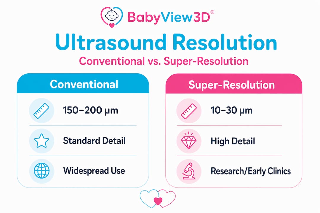
Conventional ultrasound has a hard ceiling on how fine it can resolve structures. The diffraction limit in clinical systems sits around 150 to 200 micrometers. Below that threshold, standard ultrasound simply can't tell two things apart. That's been the accepted boundary for decades.
Super-resolution ultrasound changes that entirely. By using microbubble contrast agents and a technique called ultrasound localization microscopy, researchers have pushed resolution down to as fine as 10 micrometers. That's roughly ten times sharper than what standard clinical systems can achieve today. At that scale, you can visualize individual capillaries and blood flow direction in microvessels that are completely invisible to conventional imaging.
Microbubble contrast agents create detailed maps of microvascular structures, tracking blood velocity and flow direction with extraordinary precision. Right now, this technology is primarily used in research and adult vascular imaging, but the pathway toward fetal vascular assessment is clear.
Here's how super-resolution compares to conventional ultrasound imaging:
| Feature | Conventional ultrasound | Super-resolution ultrasound |
|---|---|---|
| Spatial resolution | 150 to 200 micrometers | ~10 micrometers |
| Vessel visibility | Larger vessels only | Capillaries and microvessels |
| Blood flow mapping | Limited Doppler data | Detailed velocity and direction |
| Current clinical use | Widespread prenatal use | Research, emerging diagnostics |
| Penetration depth | Up to several centimeters | Comparable to conventional |
You can follow ultrasound trends in 2026 to stay current on where these advances are heading in prenatal care specifically.
How resolution affects your prenatal ultrasound experience
All three types of resolution, plus the frequency choices made by your sonographer, converge into the image you actually see on screen. If you've noticed that some facilities produce dramatically better pictures than others, this is why. It's not luck. It's physics, equipment calibration, and the sonographer's ability to optimize settings in real time.
In practical terms, here's what higher resolution enables during a prenatal session:
- Clear facial features during 3D imaging, including eye socket definition, nose shape, and lip profile
- Visible finger and toe separation in the second trimester
- Smooth, real-time fetal movement during 4D sessions
- Reliable measurement of small structures critical for diagnostic accuracy, such as the nuchal translucency in first-trimester screening
Adjusting focal zones, probe frequency, and sector angle can optimize image resolution even without changing the physical hardware. A skilled sonographer knows this and applies it throughout your session, not just at the start.
Resolution also connects directly to diagnostic confidence. When a sonographer can clearly see fetal anatomy, measurements are more accurate, and ambiguous findings are less common. That matters for your peace of mind just as much as it does for clinical accuracy. You can learn more about how image clarity shapes the memory-making side of prenatal scans in this piece on prenatal imaging and memories.
The type of scan also changes what resolution dimension matters most. For a 2D anatomy scan, axial and lateral resolution drive diagnostic value. For a 4D bonding session, temporal resolution steps forward because smooth motion is what makes the experience feel real and connected. And HD Live technology layers lighting simulation on top of high-resolution imaging to produce the photorealistic quality you see in premium prenatal studios.
My honest take on resolution and what actually matters
I've spent a lot of time with the technical side of prenatal ultrasound, and here's what I'd tell any expecting parent who comes to me asking which clinic has "the best resolution."
Higher resolution on paper does not automatically equal the most meaningful experience for your family. I've seen technically superior machines produce images that felt cold and clinical, while a well-calibrated system operated by a gifted sonographer produced images that made parents cry in the best possible way. Resolution is a tool. The sonographer is the artist.
What I've actually found matters more than the raw numbers is this: how well the imaging facility optimizes its equipment for your specific stage of pregnancy, how experienced the sonographer is at reading fetal position and adjusting settings, and whether the system being used combines high spatial resolution with strong temporal resolution for real-time sessions. Most parents aren't evaluating axial resolution specs. They're looking for a moment of genuine connection with their baby, and that requires all three resolution types working together at their best.
The rise of super-resolution technology is exciting, but its prenatal applications are still emerging. For right now, what separates a good prenatal imaging experience from a great one is a combination of quality equipment, skilled technique, and a facility committed to getting all the settings right before a single image is captured. That's where your energy as a parent is best spent.
— LENIER
See your baby with exceptional clarity at Bbview3d
If this article got you thinking about what your prenatal images could actually look like with the right technology behind them, Bbview3d is worth exploring.
Bbview3d has been delivering premium prenatal ultrasound experiences for over 15 years, with certified sonographers and HD Live imaging technology that combines high spatial and temporal resolution to produce the clearest, most lifelike images available today. Their 3D, 4D, and 8K sessions are specifically designed to optimize image quality for each stage of pregnancy, giving you pictures you'll keep forever. Check out their prenatal services to see package options, current promotions for first-time visitors, and studio locations across the United States.
FAQ
What does ultrasound resolution mean?
Ultrasound resolution refers to a system's ability to distinguish fine detail in an image. It includes axial resolution (depth clarity), lateral resolution (side-by-side clarity), and temporal resolution (motion clarity), all of which affect what a sonographer can see during a scan.
What factors affect ultrasound image quality?
Probe frequency, focal zone settings, scanning depth, sector angle, and the sonographer's technique all affect image quality. Higher frequency probes improve detail but reduce penetration, while adjusting scan parameters can optimize image clarity in real time.
How does frequency affect ultrasound resolution?
Higher frequencies produce sharper images but penetrate less deeply. Lower frequencies reach further into tissue but produce less detail. Sonographers select frequency based on fetal depth, which changes throughout pregnancy.
What is super-resolution ultrasound?
Super-resolution ultrasound uses microbubble contrast agents and ultrasound localization microscopy to achieve resolutions around 10 micrometers, far beyond the 150 to 200 micrometer diffraction limit of conventional systems. It's currently used in research settings with growing potential for clinical prenatal applications.
When is the best time for a high-resolution 3D ultrasound?
Between 26 and 32 weeks of pregnancy, the fetus has enough fat tissue for defined facial features and is still at a depth where frequency and penetration are well-balanced, producing the clearest and most detailed 3D images.