
Prenatal ultrasound terminology is the specialized clinical language healthcare providers use to describe your baby's development, scan findings, and imaging results during pregnancy. Explaining prenatal ultrasound terminology matters because the words in your report were written for radiologists, not for you. Terms like "echogenic," "anechoic," and "unremarkable" can feel alarming when they are actually routine descriptors. This guide decodes the most common terms, abbreviations, and report phrases you will encounter across every trimester, so you walk out of every appointment informed rather than anxious.
What are the key ultrasound terms you will encounter during prenatal scans?
Ultrasound, sonography, and sonogram are three distinct terms that parents often use interchangeably, but each means something different. Ultrasound refers to the high-frequency sound waves used to produce images, with standard pregnancy scans operating between 3 and 7.5 MHz. Sonography is the practice of performing the scan. A sonogram is the actual image produced. Knowing this distinction helps you ask better questions during appointments.
Imaging descriptors are the words that tell you what a structure looks like on screen. Here are the most common ones you will see in a prenatal report:
- Echogenic: Appears bright white on screen, typically indicating solid tissue, bone, or fat. A highly echogenic structure reflects sound waves strongly.
- Anechoic: Appears black on screen, indicating fluid-filled spaces. Amniotic fluid and the bladder are naturally anechoic.
- Hypoechoic: Appears darker than surrounding tissue, often indicating softer or less dense structures.
- Homogeneous: Uniform texture throughout, generally a reassuring descriptor for organs like the placenta.
- Heterogeneous: Mixed texture, which may prompt further review but does not automatically signal a problem.
- Unremarkable: One of the most positive words in any report. "Unremarkable" means the structure is within normal limits, with no abnormalities detected.
Anatomical terms are equally important. The gestational sac is the fluid-filled structure visible in early pregnancy, usually detectable by week 5. Crown-rump length (CRL) measures the embryo from head to bottom and is the most accurate way to confirm gestational age in the first trimester. The amniotic fluid index (AFI) measures the volume of fluid surrounding your baby, and values outside the normal range prompt closer monitoring.
Pro Tip: When you receive your report, circle every word you do not recognize before your follow-up appointment. Bring that list to your provider. A 10-minute conversation can eliminate hours of unnecessary worry.

How to understand measurement abbreviations and fetal growth indicators
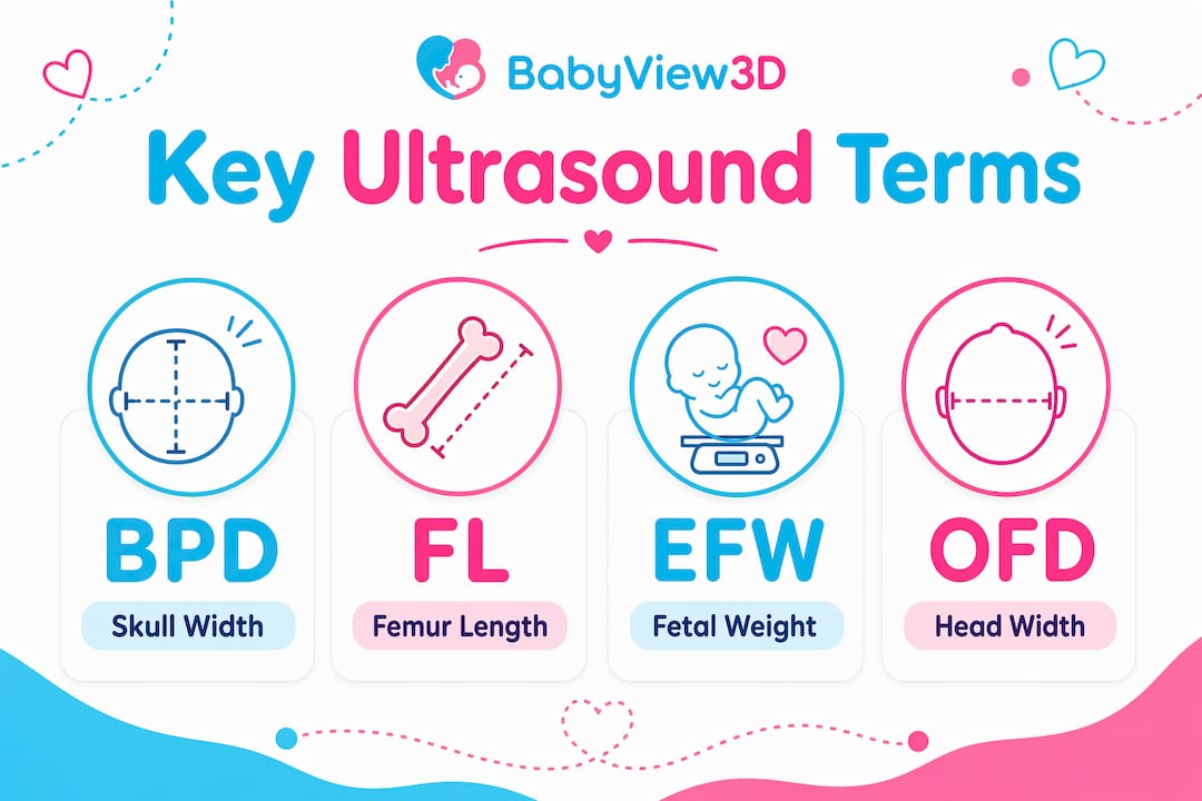
Common fetal growth indicators in ultrasound reports include BPD, FL, EFW, and OFD. These biometric parameters correlate directly with gestational age and help your provider track whether your baby is growing on schedule. Each measurement is plotted against established growth charts, and a single measurement outside the average range is rarely cause for concern on its own.
| Abbreviation | Full Term | What It Indicates |
|---|---|---|
| BPD | Biparietal diameter | Width of the baby's skull; used to estimate gestational age |
| FL | Femur length | Length of the thigh bone; tracks skeletal growth |
| EFW | Estimated fetal weight | Calculated weight based on multiple measurements combined |
| OFD | Occipitofrontal diameter | Front-to-back skull measurement; used alongside BPD |
| CRL | Crown-rump length | Head-to-bottom length; most accurate in the first trimester |
| AFI | Amniotic fluid index | Total amniotic fluid volume across four uterine quadrants |
These measurements work together, not in isolation. A provider calculating EFW combines BPD, FL, abdominal circumference (AC), and sometimes OFD into a formula. The result is an estimate, not a precise weight, with a margin of roughly 10 to 20 percent. Parents sometimes fixate on EFW as a final number, but your provider is watching the growth trend across multiple scans, not a single data point.
Pro Tip: Ask your sonographer to show you which measurement they are taking in real time. Watching BPD being measured on screen makes the abbreviation far less abstract when you read it in your report later.
Understanding normal ranges also requires context. A baby measuring slightly small for gestational age on FL alone, while all other parameters are within range, is very different from a baby showing consistently low measurements across BPD, FL, and EFW. Your provider uses the full picture to make any clinical judgment.
What do common ultrasound report phrases mean?
Ultrasound reports are written primarily for healthcare professionals using structured clinical language designed for rapid communication, not plain-language diagnosis for patients. That gap between clinical writing and patient comprehension is exactly why so many parents feel confused after reading their results. The phrases below appear in the majority of prenatal reports, and understanding them removes most of the anxiety.
- "Unremarkable": Normal. No abnormalities detected in that structure. This is the word you want to see.
- "Clinical correlation advised": The radiologist has noted something that needs to be interpreted alongside your symptoms or medical history. This phrase usually indicates a likely benign finding that requires context, not alarm.
- "Incidental finding": Something was observed that was not the focus of the scan. Most incidental findings in prenatal imaging are benign.
- "Subchorionic hemorrhage": A collection of blood between the placenta and the uterine wall. Small ones are common and often resolve without intervention.
- "Placenta previa": The placenta is positioned low in the uterus, partially or fully covering the cervix. This is monitored closely because it can affect delivery planning.
- "Mass" or "lesion": These terms denote identified areas to assess; many are ultimately benign. Neither word is a diagnosis.
The most important part of any ultrasound report is the Impression section. The Impression section condenses complex findings into a concise statement about whether results are normal or require follow-up. If you read nothing else in your report, read the Impression. It is the radiologist's summary of everything observed, written in the clearest language the report contains.
Functional ovarian cysts resolve naturally within 2 to 3 menstrual cycles, and uterine fibroids are present in up to 70 percent of women by age 50. Both are frequently noted as incidental findings in prenatal scans. Seeing either term in your report does not mean a problem has developed during your pregnancy.
What is the ultrasound procedure like and how do technical terms connect to it?
Understanding the procedure itself makes the terminology in your report feel grounded rather than abstract. Here is what happens during a standard prenatal ultrasound and what each step is actually called:
- Transducer placement. The sonographer places a handheld device called a transducer against your abdomen or, in early pregnancy, uses a transvaginal transducer for clearer images. The transducer emits and receives sound waves.
- Gel application. Ultrasound gel eliminates air between the transducer and skin, because air blocks ultrasound energy. The gel acts as a coupling medium so sound waves travel cleanly into the body.
- Real-time imaging. The machine converts returning sound waves into a live image on screen. What you see moving in real time is called a B-mode (brightness mode) scan, the standard format for prenatal imaging.
- Image capture. The sonographer freezes frames and records measurements. These saved images become your sonogram.
- 3D and 4D rendering. When advanced 3D and 4D imaging is used, the machine compiles multiple 2D slices into a surface-rendered view. 4D simply adds the dimension of real-time movement to a 3D image.
The distinction between ultrasound, sonography, and sonogram matters most here. Asking your provider "is that the procedure or the sonogram image?" during your appointment is a practical way to stay oriented. Providers sometimes use all three words loosely, and clarifying in the moment prevents confusion when you review the report later.
Technologies like HD Live, used by providers including Bbview3d, add surface-lighting algorithms to 3D rendering. This produces images with shadow and depth that look significantly more lifelike than standard 3D. The terminology you may see in reports from these sessions includes "surface rendering," "volume mode," and "HD Live mode," all of which describe how the image was processed rather than what was found clinically.
Key takeaways
Prenatal ultrasound reports use clinical language built for radiologists, and knowing the core terms transforms a confusing document into a clear picture of your baby's development.
| Point | Details |
|---|---|
| "Unremarkable" is positive | This term means the structure is normal with no abnormalities detected. |
| Read the Impression section first | It summarizes all findings in the clearest language the report contains. |
| Measurements are trends, not verdicts | BPD, FL, EFW, and OFD are tracked across scans, not judged as single data points. |
| Cautious phrases are precautionary | "Clinical correlation advised" usually signals a benign finding needing context, not a diagnosis. |
| Ask during the scan | Clarifying terms in real time with your sonographer prevents misreading the report later. |
What parents actually get wrong about ultrasound reports
Most parents I speak with make the same mistake: they read the body of the report before the Impression, get stopped by a phrase like "heterogeneous echotexture" or "echogenic focus," and spend the next 48 hours convinced something is seriously wrong. The Impression is at the bottom of the page for structural reasons, not because it is less important. It is the most important sentence in the document. Read it first, every time.
The second mistake is treating measurement percentiles the way parents treat school grades. A femur length at the 15th percentile is not a failing grade. It is one data point in a population of normal variation. Providers look for consistent patterns across multiple measurements and multiple appointments. One number sitting outside the middle range rarely changes clinical management.
What I find genuinely reassuring is that radiologists use cautious phrasing like "monitoring recommended" precisely because they are being careful, not because they have found something alarming. That caution is the system working correctly. If you see follow-up language in your report, treat it as your provider doing their job thoroughly, not as a warning sign.
Keep a simple glossary on your phone. Write down every term you encounter across your scans. By your third trimester, you will have built enough fluency to read your own reports with confidence. That fluency does not replace your provider's judgment, but it makes every conversation with them more productive.
— LENIER
See your baby clearly with Bbview3d
Bbview3d has spent over 15 years helping expectant parents see their babies in extraordinary detail, using HD Live, 3D, 4D, and 8K imaging technology at centers across the United States. Every session is guided by certified sonographers who explain what you are seeing in plain language, so the terminology in your report connects directly to the face on the screen. When you understand what you are looking at, the experience becomes something far more than a medical appointment. Explore prenatal imaging packages designed to give your family the clearest possible view of your baby before birth, with a first-appointment discount available for new families.
FAQ
What does "unremarkable" mean in a prenatal ultrasound report?
"Unremarkable" is a positive term meaning the structure examined shows no abnormalities and is within normal limits. It is one of the best phrases to find in your report.
What is the difference between ultrasound, sonography, and sonogram?
Ultrasound refers to the high-frequency sound waves used in imaging, sonography is the practice of performing the scan, and a sonogram is the actual image produced during the session.
What do BPD, FL, and EFW stand for in my report?
BPD is biparietal diameter (skull width), FL is femur length, and EFW is estimated fetal weight. These biometric measurements are used together to track fetal growth and confirm gestational age.
Should I be worried if my report says "clinical correlation advised"?
This phrase typically means a finding was noted that needs to be interpreted alongside your symptoms or medical history. It is usually precautionary and does not indicate a serious problem.
Which part of the ultrasound report should I read first?
Read the Impression section first. It condenses all findings into a clear summary statement and tells you directly whether results are normal or require follow-up.