

A sonogram session is a non-invasive imaging procedure that uses high-frequency sound waves to produce live pictures of the inside of your body. The terms "sonogram" and "ultrasound" are used interchangeably by medical professionals and patients alike. Both refer to the same safe, radiation-free process. For expectant parents, understanding what does a sonogram session involve removes anxiety and turns a clinical appointment into one of the most memorable moments of pregnancy.
Ultrasound technology is radiation-free, making it safe at every stage of pregnancy. A standard session lasts between 15 and 60 minutes, depending on the type of exam and what the sonographer needs to assess. You watch the images appear on a monitor in real time. That live view of your baby is what makes the sonogram appointment process so emotionally powerful for families.
What does a sonogram session involve before you arrive?
Preparation for a sonogram varies based on the type of exam scheduled. Some pelvic and early pregnancy scans require a comfortably full bladder because a full bladder pushes the uterus upward, giving the sonographer a clearer view. Other scans, including most second and third trimester appointments, require no special preparation at all.
Here is what to plan for on the day of your appointment:
- Arrive 15 minutes early. Most facilities ask you to complete intake paperwork before the session begins. Arriving late compresses your actual scan time.
- Wear comfortable, two-piece clothing. The sonographer needs easy access to your abdomen. Loose pants and a top you can lift are ideal.
- Bring your referral and insurance information. Some imaging centers require a physician's order before starting the exam.
- Drink water beforehand if instructed. For early pregnancy pelvic scans, your provider will tell you exactly how much to drink and when to stop urinating.
- Bring a support person. Most facilities allow one or two guests. Confirm the policy when you book.
Pro Tip: Call the imaging center the day before to confirm your specific preparation instructions. Requirements differ between a first trimester dating scan and a 20-week anatomy scan, so never assume the same rules apply.
Step-by-step walkthrough of the sonogram procedure
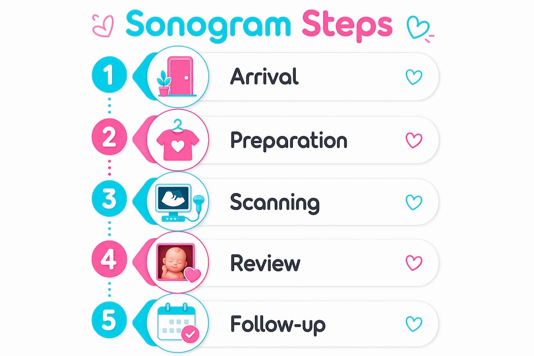
Knowing the sonogram session steps in order makes the experience feel familiar rather than clinical. Here is exactly how a standard abdominal sonogram unfolds from start to finish.
- You lie on the exam table. The sonographer positions you on your back, usually with a pillow under your head. The table is padded and the room is dimly lit so the monitor is easier to read.
- The sonographer applies conductive gel. The gel is water-soluble and non-staining, but it tends to feel cold on contact. Some clinics warm the gel to improve comfort, especially during longer sessions.
- The transducer moves across your skin. The sonographer presses a small handheld device called a transducer against your abdomen and glides it in different directions. The transducer sends sound waves into your body and picks up the echoes that bounce back. Those echoes become the real-time images you see on the monitor.
- You may be asked to hold your breath or shift position. Patient cooperation directly impacts image quality. Holding your breath for a few seconds displaces bowel gas and steadies the image. A slight turn to one side can improve the angle on the baby's spine or heart.
- The sonographer captures still images and measurements. Throughout the scan, the sonographer freezes frames and records measurements of the baby's head, abdomen, and femur. These numbers feed directly into your prenatal care record.
- The gel is wiped away. Once the scan is complete, the sonographer hands you a towel or paper wipe to remove the gel. The entire process is painless and leaves no residue on your clothing.
Pro Tip: Ask the sonographer to narrate what they are seeing as they scan. Most are happy to point out the baby's heartbeat, limbs, and face in real time. It transforms a medical procedure into a genuine bonding moment.
What are the different types of sonograms during pregnancy?

Not every sonogram appointment process looks the same. The type of scan your provider orders depends on how far along you are and what clinical information is needed.
| Sonogram type | When it is used | What it shows |
|---|---|---|
| Transabdominal | Second and third trimester | Fetal anatomy, growth, placenta position |
| Transvaginal | First trimester, early pregnancy | Heartbeat confirmation, gestational sac |
| 2D ultrasound | Any stage | Standard black and white cross-section images |
| 3D ultrasound | 26–32 weeks optimal | Surface rendering of baby's face and body |
| 4D ultrasound | 26–32 weeks optimal | Live motion video of baby's movements |
Transvaginal sonograms use a sterile covered probe placed internally, which some patients find unfamiliar. The procedure is safe and typically brief. It is the most reliable method for confirming a heartbeat before 10 weeks because the probe sits closer to the uterus than an abdominal transducer can reach.
The jump from 2D to 3D and 4D imaging is significant for families. A 2D scan gives your provider the clinical measurements they need. A 3D ultrasound session adds surface detail that lets you recognize your baby's nose, lips, and cheeks before birth. A 4D scan adds motion, so you watch your baby yawn, stretch, or suck their thumb in real time. The emotional benefits of prenatal imaging at this level go well beyond clinical data. Many families describe 3D and 4D sessions as the moment pregnancy became fully real to them.
What happens after the sonogram session ends?
Understanding the follow-up process is just as important as knowing how the scan works. Here is what to expect once the session is complete.
- Real-time viewing during the scan. You see the images as they are captured. The sonographer may explain what they are measuring, though a full clinical interpretation comes later.
- Radiologist review. After the session, a radiologist studies the captured images and measurements in detail. Results are typically sent to your healthcare provider within 1–2 days.
- Your provider discusses the findings. Your OB, midwife, or maternal-fetal medicine specialist reviews the radiologist's report and contacts you to discuss results. Never rely solely on what the sonographer says during the scan as a final clinical verdict.
- Follow-up scans may be scheduled. If the baby's position blocked a key measurement or if a finding needs monitoring, your provider will order a repeat scan. This is common and does not automatically signal a problem.
- Keepsake images are available at some centers. Elective imaging centers like Bbview3d offer printed photos, digital files, and video recordings of the session. These are separate from the clinical report and are yours to keep.
The gap between the session and your results conversation can feel long. Write down any questions you have during the scan and bring them to your follow-up appointment. Your provider is the right person to interpret what the images mean for your specific pregnancy.
Key takeaways
A sonogram session is a safe, painless procedure that uses sound waves to produce real-time images, typically lasting 15–60 minutes, with preparation, procedure type, and follow-up steps varying by trimester and clinical need.
| Point | Details |
|---|---|
| Preparation varies by scan type | Full bladder required for some early scans; confirm instructions the day before your appointment. |
| Gel and transducer are the core tools | Water-soluble gel and a handheld transducer create real-time images with no radiation exposure. |
| Cooperation improves image quality | Holding your breath and shifting position when asked gives the sonographer clearer, more accurate images. |
| Results take 1–2 days | A radiologist reviews images after the session and sends a report to your healthcare provider. |
| 3D and 4D scans add emotional depth | Surface-rendering technology lets families see facial features and live movement beyond standard clinical imaging. |
Why the sonogram experience matters more than most parents expect
I have spent years watching families walk into sonogram sessions tense and walk out transformed. The clinical side of the procedure is straightforward. What surprises people is how much the experience itself shapes their connection to the pregnancy.
The single most underused tool in any sonogram appointment is your voice. Ask the sonographer to explain what they are looking at. Ask them to show you the heartbeat on the screen. Ask what each measurement means. Sonographers are trained clinicians, not tour guides, but most genuinely enjoy sharing what they see when a parent is engaged. The session becomes collaborative rather than passive.
I also think parents underestimate how much their own stillness matters. Patient cooperation significantly impacts image quality. A blurry image is not just a technical inconvenience. It can mean a repeat appointment, more waiting, and more anxiety. Staying still and following breath instructions is one of the few things you can actively control in the process.
The emotional weight of seeing your baby's face in 3D for the first time is something no preparation fully captures. I have seen parents cry, laugh, and go completely silent. All of those reactions are right. What I would tell any expectant family is this: let yourself be present in that room. The images on the screen are not just data. They are your first real introduction to your child.
— LENIER
See your baby in stunning detail with Bbview3d
Bbview3d has specialized in prenatal ultrasound imaging for over 15 years, with certified sonographers and advanced 3D, 4D, and HD Live technology across multiple locations in the United States. If a standard clinical scan leaves you wanting more than a blurry black and white printout, Bbview3d offers premium imaging packages designed specifically for expectant families. You get high-resolution images, video recordings, and keepsakes that capture your baby's features in remarkable detail. Explore the full range of session packages and book your first appointment, often available at a discounted rate for new families.
FAQ
What does a sonogram session involve from start to finish?
A sonogram session involves lying on an exam table while a sonographer applies conductive gel and moves a transducer across your skin to produce real-time images. The full process, including paperwork and the scan itself, typically takes 15 to 60 minutes.
Do I need to prepare differently for each type of sonogram?
Yes. Some early pregnancy and pelvic scans require a full bladder for clearer imaging, while most second and third trimester scans need no special preparation. Always confirm your specific instructions with the imaging center when you book.
Is a sonogram safe during pregnancy?
Sonograms are radiation-free and safe throughout all stages of pregnancy. They use sound waves rather than ionizing radiation, which is why they are the standard imaging tool for fetal monitoring.
How long does it take to get sonogram results?
A radiologist typically reviews the images and sends a report to your healthcare provider within 1–2 days. Your provider then contacts you to discuss the findings and any recommended next steps.
What is the difference between a 2D and a 3D sonogram?
A 2D sonogram produces flat, cross-section images used for clinical measurements and fetal assessment. A 3D sonogram renders surface detail, showing your baby's facial features and body contours in a way that 2D imaging cannot capture.