

A first prenatal ultrasound is a medical imaging procedure that uses sound waves to create pictures of the developing embryo inside the uterus, confirming pregnancy viability, estimating gestational age, and detecting multiple pregnancies. Clinically called an obstetric ultrasound, this scan is the first concrete proof that a pregnancy is progressing as expected. Most providers schedule it between 6 and 10 weeks of pregnancy. For expectant parents, it is also the first time they see and hear their baby, making it one of the most emotionally charged appointments of the entire pregnancy.
What is a first prenatal ultrasound like, and when does it happen?
The timing of your first prenatal ultrasound depends on your provider, but most scans happen between 6 and 9 weeks of pregnancy. Earlier scans confirm viability and rule out complications like ectopic pregnancy. Later scans in the first trimester, around 11–14 weeks, add early screening for structural concerns.
Two types of ultrasound are used in early pregnancy, and which one you receive depends on how far along you are:
- Transvaginal ultrasound: Used before 10–12 weeks. A slender, lubricated probe is gently inserted into the vagina to capture images. Transvaginal ultrasound provides clearer images early in pregnancy because of its proximity to the uterus, especially before the embryo is visible through the abdomen.
- Abdominal ultrasound: Used after 12 weeks. A handheld transducer glides across your lower abdomen with gel applied to the skin. This is the version most parents picture when they imagine an ultrasound.
Both types are safe and non-invasive, with no increased risk of miscarriage. The transvaginal probe may feel unfamiliar, but it does not cause pain. Knowing this in advance removes most of the anxiety around the procedure.
Pro Tip: Ask your provider in advance which type of ultrasound to expect. If you are having a transvaginal scan, wearing a skirt or loose pants makes the preparation faster and more comfortable.
What happens step by step during the appointment?
The appointment itself is straightforward. Most first-trimester ultrasound scans last between 20 and 45 minutes, depending on how cooperative the embryo is and what the sonographer needs to measure.
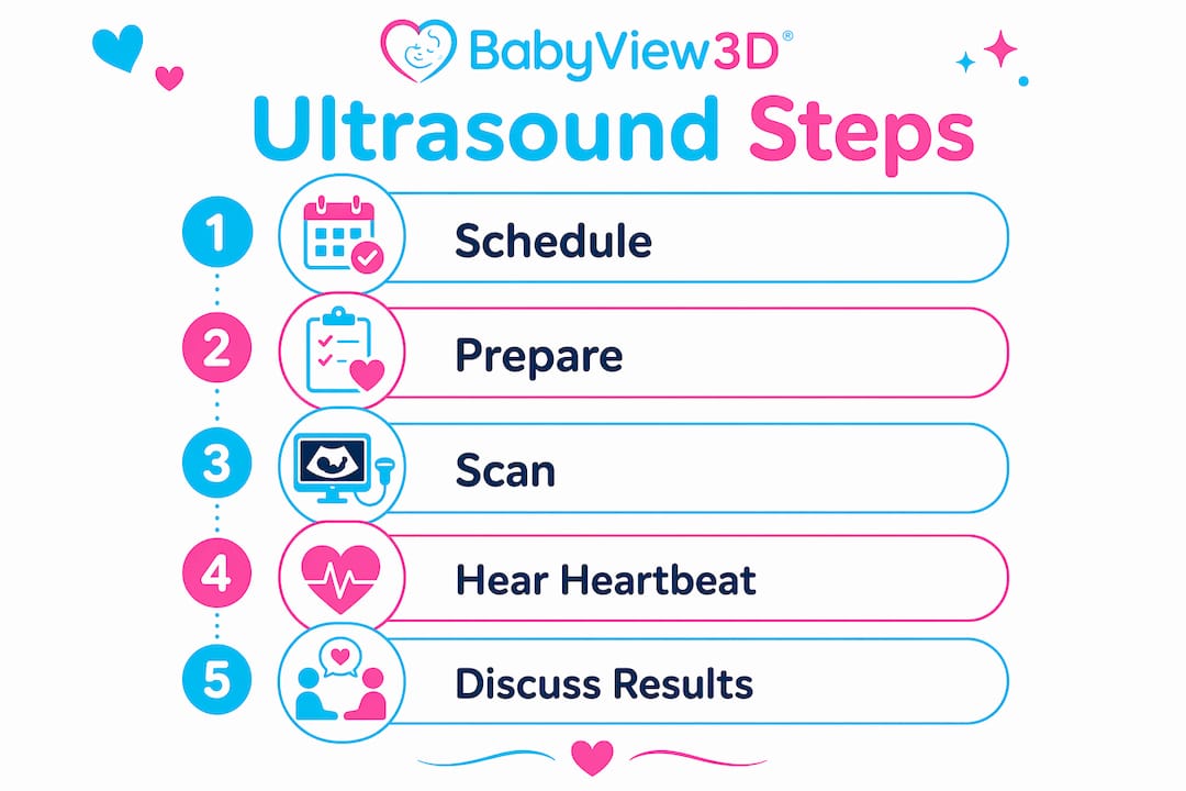
Here is what typically happens, in order:
- Preparation: For an abdominal ultrasound, you may be asked to arrive with a full bladder, since a full bladder pushes the uterus into a better imaging position. For a transvaginal scan, a full bladder is not required. Following your provider's bladder instructions and wearing comfortable clothing makes the process easier.
- Positioning: You lie on an exam table. For abdominal scans, gel is applied to your lower abdomen. For transvaginal scans, the probe is covered with a sterile sheath and lubricated before insertion.
- Imaging: The sonographer moves the probe to capture images from multiple angles. You will see a grainy black-and-white image on a monitor. The embryo at 6–8 weeks is tiny, roughly the size of a blueberry, so the image may look like a small bright spot.
- Measurements: The sonographer takes the crown-rump length (CRL), which is the longest straight-line measurement of the embryo. CRL is the primary tool for calculating gestational age between 6 and 10 weeks.
- Heartbeat check: The sonographer confirms a fetal heartbeat. This is the moment most parents describe as the most emotional part of the entire scan.
- Results discussion: Your provider reviews findings with you, including your estimated due date and any immediate observations.
The sonographer also looks for the yolk sac inside the gestational sac. The yolk sac is an early visual marker that confirms the pregnancy is intrauterine, meaning it is developing inside the uterus and not in a fallopian tube.
Pro Tip: Bring a partner, family member, or close friend to the appointment. Sharing the moment of hearing the heartbeat for the first time makes the experience more meaningful, and having someone with you helps if emotions run high.

Why does the first ultrasound feel so emotional?
Hearing the baby's heartbeat for the first time is consistently described as the most exciting and emotional moment of the first prenatal ultrasound. The rapid flutter of a heartbeat at 6–8 weeks, often between 90 and 170 beats per minute, makes the pregnancy feel suddenly real in a way that a positive test strip cannot.
Expectant parents experience a wide range of feelings during this appointment:
- Joy and relief: Seeing a heartbeat confirms the pregnancy is progressing.
- Anxiety: Many parents feel nervous before the scan, especially if they have experienced a previous loss.
- Surprise: The embryo is much smaller than most parents expect. Understanding that fetal size at this stage is tiny helps manage expectations before you walk into the room.
- Bonding: Seeing the embryo on screen, even as a small bright shape, triggers a powerful emotional connection for many parents.
All of these reactions are normal. Practitioners who perform early prenatal scans regularly report that the emotional range in the room, from tears of joy to quiet shock, is part of the experience.
"Hearing the heartbeat is often described as the most exciting moment and emotional highlight for expectant parents. The emotional spectrum during the initial scan is normal and expected."
Learning what to expect before your appointment, including the small size of the embryo and the grainy quality of early images, reduces anxiety and lets you be present for the emotional experience. Reading about the emotional impact of ultrasound before your appointment gives you a realistic picture of what the moment feels like.
Why does the first ultrasound matter clinically?
The first prenatal ultrasound does more than confirm a pregnancy exists. It collects data that shapes your entire prenatal care plan.
| Clinical Goal | What the Sonographer Checks | Why It Matters |
|---|---|---|
| Confirm intrauterine location | Gestational sac and yolk sac position | Rules out ectopic pregnancy |
| Gestational dating | Crown-rump length (CRL) measurement | Sets accurate due date |
| Viability | Fetal heartbeat presence | Confirms pregnancy is progressing |
| Multiple pregnancies | Number of gestational sacs | Identifies twins or higher-order multiples |
| Early anomaly screening | Nuchal translucency at 11–14 weeks | Flags potential chromosomal concerns |
Ultrasound is the gold standard for accurate gestational dating, often correcting discrepancies with last menstrual period calculations. This matters because your due date determines the timing of every subsequent prenatal test and scan.
The first-trimester anatomy scan, performed around 11–14 weeks, adds another layer of clinical value. This scan can detect 40–70% of major structural anomalies, with detection rates improving closer to 13–14 weeks. It does not replace the detailed anatomy scan performed around 20 weeks, but it provides early information that can guide further testing.
Detailed first-trimester ultrasound exams require specialized equipment and trained sonographers. Guidelines from the International Society of Ultrasound in Obstetrics and Gynecology (ISUOG) and the American Institute of Ultrasound in Medicine (AIUM) increasingly recommend offering these detailed scans where equipment and expertise allow. The quality of your first scan depends directly on the skill of the practitioner and the technology available.
While clinicians focus on confirming date, location, and pregnancy health, parents focus on the visual and auditory experience. Both perspectives are valid. Understanding what clinicians look for helps you ask better questions and understand the results you receive.
Key Takeaways
The first prenatal ultrasound is a safe, non-invasive scan that confirms viability, establishes gestational age, and delivers the first emotional milestone of pregnancy for expectant parents.
| Point | Details |
|---|---|
| Timing matters | Most first scans happen between 6 and 10 weeks, with 11–14 weeks adding early anomaly screening. |
| Two scan types | Transvaginal ultrasound is used before 10–12 weeks; abdominal ultrasound is used after 12 weeks. |
| CRL is the key measurement | Crown-rump length sets your due date more accurately than last menstrual period dating alone. |
| Emotional range is normal | Joy, anxiety, and surprise are all common reactions during the first ultrasound appointment. |
| Skill and equipment matter | Detailed first-trimester scans require trained sonographers and advanced imaging technology. |
What I've learned from watching parents experience their first scan
I have spent years observing how expectant parents prepare for, and respond to, the first prenatal ultrasound. The single biggest factor in how well the experience goes is preparation. Parents who understand the procedure beforehand, including why the image looks grainy, why the embryo is so small, and why the transvaginal probe is used, walk out of the room feeling informed rather than confused.
The parents who struggle most are the ones who arrive expecting a clear, detailed image of a recognizable baby. At 7 weeks, the embryo looks like a small curved shape on a dark screen. That is not a failure of the technology. That is biology. Setting that expectation before the appointment changes everything.
The heartbeat is where the emotional shift happens for almost everyone. No amount of preparation fully prepares you for that sound. My honest observation is that the first ultrasound is less about what you see and more about what you hear. That rapid flutter confirms that something remarkable is happening inside the body, and no amount of reading about it substitutes for the moment itself.
Preparation reduces anxiety. Knowledge creates space for emotion. Go in informed, and let yourself feel whatever comes next.
— LENIER
Bbview3d: a closer look at your baby, earlier
Knowing what to expect is one thing. Seeing your baby in detail is another experience entirely.

Bbview3d has over 15 years of experience helping expectant parents see their babies with clarity and warmth. With certified sonographers and advanced 3D, 4D, and 8K HD Live imaging technology, Bbview3d creates a comfortable, family-centered environment where the first visual encounter with your baby becomes a memory you keep. Explore prenatal imaging packages designed for every stage of pregnancy, including early sessions for parents who want a detailed first look. First-appointment discounts are available for new families. Visit Bbview3d to see what is possible.
FAQ
When is the first prenatal ultrasound scheduled?
Most providers schedule the first prenatal ultrasound between 6 and 10 weeks of pregnancy to confirm viability and estimate the due date.
Does the transvaginal ultrasound hurt?
The transvaginal probe may feel unfamiliar but is not painful. The probe is sterile and lubricated, and most parents report only mild pressure during the procedure.
What is crown-rump length and why does it matter?
Crown-rump length (CRL) is the longest straight-line measurement of the embryo and is the primary method for gestational dating between 6 and 10 weeks, often more accurate than last menstrual period calculations.
Why does the first ultrasound image look so unclear?
Early ultrasound images are grainy because the embryo is very small at 6–8 weeks. Fetal size at this stage is roughly comparable to a blueberry, so the clinical focus is on heartbeat and location rather than detailed visuals.
Can the first ultrasound detect twins?
Yes. The sonographer checks the number of gestational sacs during the first scan, making it the earliest point at which a multiple pregnancy is typically confirmed.