

First ultrasound images surprise parents because they look nothing like the clear, detailed baby photos most expectant parents picture in their minds. The clinical term for this early scan is a dating scan or viability ultrasound, and its primary job is medical measurement, not memory making. Grayscale sound waves, unexpected fetal activity, and the quirks of 3D rendering software all combine to create an experience that is equal parts confusing and deeply moving. Understanding what you are actually looking at, and why, turns that initial shock into something you will want to hold onto forever.
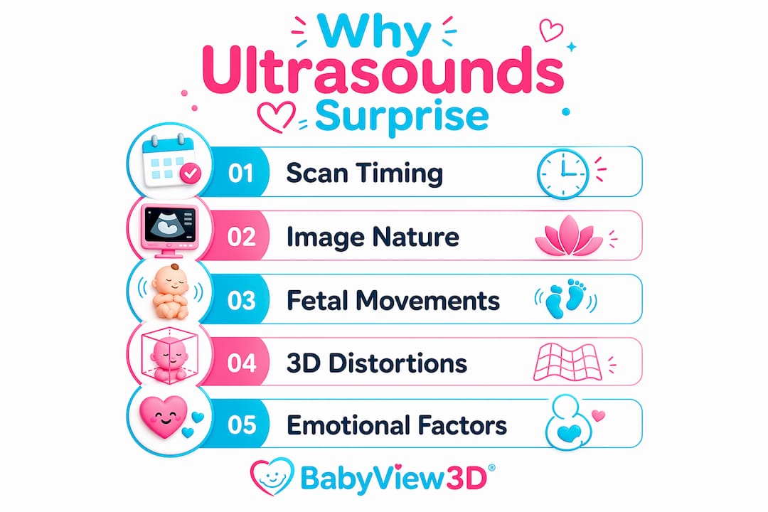
Why first ultrasound images surprise parents: timing is everything
The single biggest reason early scans look unfamiliar is when they happen. First prenatal ultrasounds are typically scheduled between 8 and 10 weeks to give the sonographer the clearest possible window for viability assessment and accurate gestational age measurement. Before 8 weeks, the embryo is often too small for consistent evaluation.
At 8–10 weeks, the embryo resembles a small "gummy bear" shape, not a recognizable baby. That gap between expectation and reality is the source of most first-time parents' surprise. Many parents walk in expecting something close to a newborn photo and walk out staring at a grainy bean shape on a printout.

The emotional peak at this stage is almost always the heartbeat, not the image. Hearing or seeing that rapid flutter on screen hits differently than any visual detail. Sonographers know this and will often pause to let you absorb it.
Pro Tip: Ask your sonographer to point out the heartbeat on screen before they move on to measurements. That single moment anchors the whole experience.
Key facts about timing and what it means for parents:
- Scans before 8 weeks may require a transvaginal probe for clearer images, which surprises many parents who expected an abdominal scan.
- The embryo measures roughly 1.6–2.5 cm at 8–10 weeks, making fine detail impossible to see.
- Sonographers prioritize crown-rump length and cardiac activity over image aesthetics at this stage.
- Earlier scans, done around 6–7 weeks, sometimes fail to detect a heartbeat yet, which can cause unnecessary alarm. Scheduling at 8–10 weeks reduces that risk significantly.
Why do ultrasound images look so confusing on screen?
Ultrasound imaging produces grayscale, sound-wave-based renderings, not photographs. The machine sends high-frequency sound waves into tissue and maps how they bounce back. Dense tissue appears white, fluid appears black, and everything in between shows up as shades of gray. That is a fundamentally different visual language than anything parents have seen before.
Sonographers focus on viability, heartbeat, and gestational age rather than creating picture-perfect images. The scan is a diagnostic tool first. Parents are not expected to identify anatomy on their own, and the sonographer's silence during measurement is normal. It reflects concentration, not concern.
Parents expecting clear photos based on pregnancy books often feel disappointed when the screen shows a blurry, shifting gray image. That disappointment fades quickly once the sonographer explains what each shadow and shape represents. For a deeper look at what affects image quality, the factors behind ultrasound clarity are worth reviewing before your appointment.
What makes early images hard to read at a glance:
- Amniotic fluid appears as dark, empty space, which can make the embryo look isolated or small.
- Shadows from the uterine wall and placenta create dark patches that look like voids.
- The embryo's position changes constantly, so the image shifts during the scan.
- Bone structures reflect sound strongly and appear bright white, while soft tissue is much harder to distinguish.
Pro Tip: Bring a partner or support person who can help you remember what the sonographer explains. You will be processing a lot of emotion and detail at the same time.
What surprising fetal movements can parents see in early scans?
Fetal activity visible on ultrasound is one of the most unexpected and emotionally powerful parts of the experience. Most parents expect a still image. What they often get is a tiny figure that moves, stretches, and responds.
Fetal physiological functions such as urination begin by weeks 10–12, which surprises nearly every parent who witnesses it. The fetus is not just growing. It is actively practicing the biological functions it will need after birth. Watching that happen in real time changes how parents think about what is happening inside the womb.
Fetal movements such as yawning, swallowing, and breathing motions are normal developmental markers that deepen parental connection when observed. These behaviors are not random. They are signs of a nervous system that is already working. For a week-by-week breakdown of what to expect, the fetal development milestones guide gives parents a clear reference point.
Here is what parents commonly see moving on early scans, and what each action means:
- Limb movements. Arms and legs begin moving by 8–10 weeks. The movements look jerky and uncoordinated, which is completely normal at this stage of muscle development.
- Swallowing. The fetus swallows amniotic fluid to practice digestive function. You may see the jaw moving rhythmically on screen.
- Yawning. Visible as early as 11–12 weeks, yawning is linked to brain development and is one of the most recognizable and emotionally striking things parents see.
- Breathing motions. The chest rises and falls as the fetus practices the mechanics of breathing, even though the lungs are not yet functional for air.
- Urination. Fetal kidneys begin filtering fluid by weeks 10–12. The resulting urine becomes part of the amniotic fluid, completing a loop that keeps the environment healthy.
Why do 3D ultrasound images sometimes look distorted?
3D ultrasound images use rendering software to reconstruct a surface image from thousands of 2D sound-wave slices. The result depends entirely on what the software has to work with. When conditions are ideal, the image is striking. When they are not, the result can look genuinely strange.
3D ultrasound distortions occur because the rendering algorithm fills in data gaps caused by obstructions like the placenta, fetal limbs, or the uterine wall. The software makes its best guess about what is behind an obstruction. Those guesses produce the unusual features that alarm parents. The algorithm can misinterpret physical obstructions like the umbilical cord as facial features, creating what looks like extra bumps or distorted cheeks.
Clear 3D images require at least 1–2 cm of amniotic fluid between the probe and the baby's face. Without that fluid buffer, the image compresses and the baby's features appear flattened or smeared. Fetal position matters just as much as fluid levels.
| Condition | Effect on 3D image |
|---|---|
| Baby facing the probe with clear fluid | Sharp, detailed facial features |
| Baby pressed against the placenta | Flattened or smeared appearance |
| Hands or cord near the face | Distorted features, extra bumps |
| Low amniotic fluid volume | Compressed, unclear rendering |
| Baby in "boxer pose" with fists up | Unusual facial contours from algorithm fill-in |
Pro Tip: Drink 32 oz of water in the two hours before a 3D scan. Better hydration increases amniotic fluid clarity and gives the rendering software more to work with.
Understanding how 3D ultrasound imaging works step by step before your session removes most of the surprise from distorted images. When you know why the software struggles, a strange-looking image stops being alarming and starts being interesting.
Key Takeaways
First ultrasound images surprise parents most because the scans are diagnostic tools, not portrait sessions, and the fetus looks and behaves in ways that no pregnancy book fully prepares you for.
| Point | Details |
|---|---|
| Timing shapes what you see | Scans at 8–10 weeks show a small embryo, not a formed baby. |
| Images are sound maps, not photos | Grayscale renderings require a sonographer's guidance to interpret. |
| Fetal activity starts early | Yawning, swallowing, and urination begin by weeks 10–12. |
| 3D distortions have technical causes | Obstructions and low fluid lead to algorithm fill-in errors. |
| The heartbeat is the real moment | Most parents find the heartbeat more moving than any visual image. |
What I have learned from watching parents see their baby for the first time
The reaction I see most often is not joy or tears. It is confusion followed by a question: "Is that normal?" Parents arrive with a mental image built from social media, pregnancy apps, and friends' keepsake photos. The actual scan rarely matches any of it. That gap is not a failure of the technology or the appointment. It is a feature of how early pregnancy actually works.
What I have noticed is that parents who understand the "why" behind the image connect faster. When you know the embryo is supposed to look like a gummy bear at 8 weeks, you stop trying to find a nose and start listening for the heartbeat instead. That shift in focus changes the whole emotional experience of the appointment.
The parents who leave most satisfied are the ones who came in curious rather than expectant. They asked questions. They let the sonographer walk them through the screen. They did not measure the appointment against a photo they saw on Instagram. The emotional benefits of prenatal imaging are real, but they require you to meet the technology where it is, not where you imagined it would be.
My honest advice: go in knowing the image will be imperfect. The imperfection is proof that something real is happening. A blurry, shifting, confusing image of a tiny moving shape is one of the most extraordinary things you will ever see.
— LENIER
What Bbview3d offers parents who want more from their scan

Bbview3d has spent more than 15 years helping families see their baby before birth with a level of detail that standard clinical scans cannot provide. Using HD Live technology alongside 3D, 4D, and 8K resolution imaging, Bbview3d's certified sonographers create sessions designed around the emotional experience, not just the medical data. The difference shows up in the image quality and in how parents feel walking out the door.
If you want to see facial features, catch a yawn, or simply understand what you are looking at, the prenatal imaging packages at Bbview3d are built for exactly that. First-appointment discounts are available, and locations across the United States make it accessible for most families.
FAQ
Why does my first ultrasound look like a blur?
Early ultrasounds are grayscale sound-wave maps, not photographs. The sonographer focuses on medical measurements, so image clarity is secondary to diagnostic accuracy.
When is the best time for a first ultrasound?
The 8–10 week window is the standard for viability and gestational age assessment. Before 8 weeks, the embryo is often too small for consistent evaluation.
Why does my baby look strange in a 3D ultrasound?
3D rendering software fills in data gaps caused by obstructions like the placenta or fetal hands. The result can look distorted, but it reflects a technical limitation, not a problem with the baby.
Can I see my baby move at the first ultrasound?
Yes. Limb movements are often visible by 8–10 weeks. Yawning, swallowing, and breathing motions appear by 11–12 weeks and are normal signs of healthy development.
Why was the sonographer so quiet during my scan?
Sonographers concentrate on measurements during the scan. Silence reflects focus, not concern. Ask them to narrate what they see once the measurement phase is complete.