

Ultrasound image quality is defined as the combined clarity, sharpness, and diagnostic usefulness of prenatal scan images produced by sound wave reflection. For expectant parents, understanding what drives that quality transforms a confusing or disappointing scan into a meaningful experience. Three forces control what you see on screen: the sonographer's machine settings, the physics of sound traveling through tissue, and your baby's position in the womb. Gain, focal zones, and probe frequency are the core technical levers. Recent AI-based evaluation frameworks and comparative studies of handheld devices have added new precision to how professionals assess and improve prenatal image clarity.
What technical factors control ultrasound image quality?
Explaining ultrasound image quality starts with the controls a sonographer adjusts before and during your scan. These settings directly determine whether your baby's face appears sharp and detailed or grainy and washed out.
Gain controls how bright the overall image appears. Think of it like the volume knob on a speaker. Turn it too high and the image floods with white noise. Turn it too low and everything goes dark. High gain settings are the most common technical cause of grainy or washed-out 3D ultrasound images, which means the first fix a skilled sonographer reaches for is always gain reduction, not a new machine.
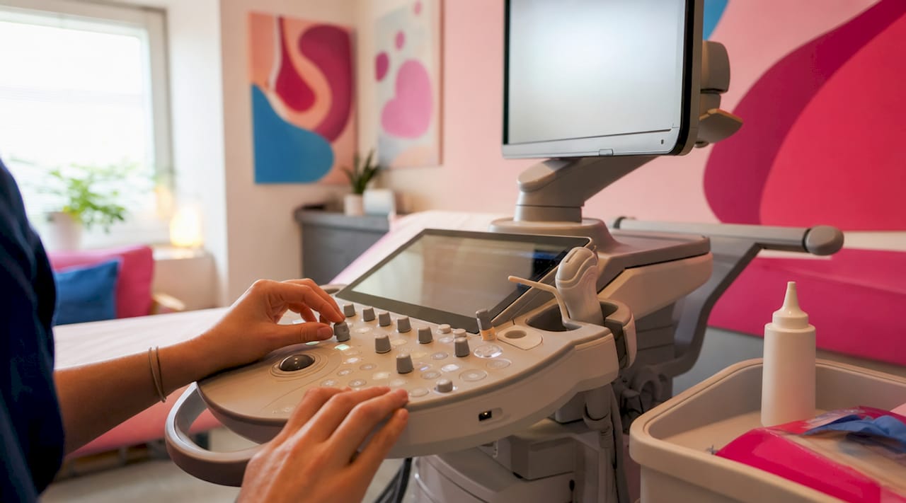
Focal zones determine where the beam sharpens its focus inside your body. Placing the focal zone at the wrong depth is like taking a photo with the autofocus locked on the background. Incorrect focal zone placement causes blurring by focusing sharpness away from the target structure, and many operators overlook this adjustment entirely. Moving the focal zone to match the depth of the baby's face can transform a blurry image into a clear one within seconds.
Probe frequency introduces a physics trade-off that every sonographer manages in real time. Increasing frequency increases resolution but decreases penetration, so a high-frequency probe gives finer detail for shallow structures while a lower frequency is needed to reach deeper ones. For a baby positioned far from the probe, the sonographer must drop frequency to maintain penetration, which slightly reduces fine detail.
Using multiple focal zones improves spatial resolution across a wider depth range, but multiple focal zones decrease frame rate, causing choppy motion in 4D video when your baby is active. This is a genuine trade-off, not a malfunction. For still 3D images, multiple zones work well. For smooth live video, a single zone at the right depth performs better.
- Reduce gain first if the image looks grainy or overexposed.
- Reposition the focal zone to match the depth of the baby's face.
- Increase smoothing to reduce speckle noise without losing structural detail.
- Adjust probe angle to find a cleaner acoustic path to the target.
- Reconsider probe frequency if depth is limiting penetration.
Pro Tip: Ask your sonographer to walk you through one setting adjustment during the session. Watching gain or focal zone change in real time makes the whole process click, and most sonographers are happy to explain what they are doing.
How do physical and anatomical factors affect ultrasound image clarity?
Technical skill and equipment quality only go so far. The single biggest variable in prenatal ultrasound image clarity is often something neither you nor the sonographer can control: your baby's position.
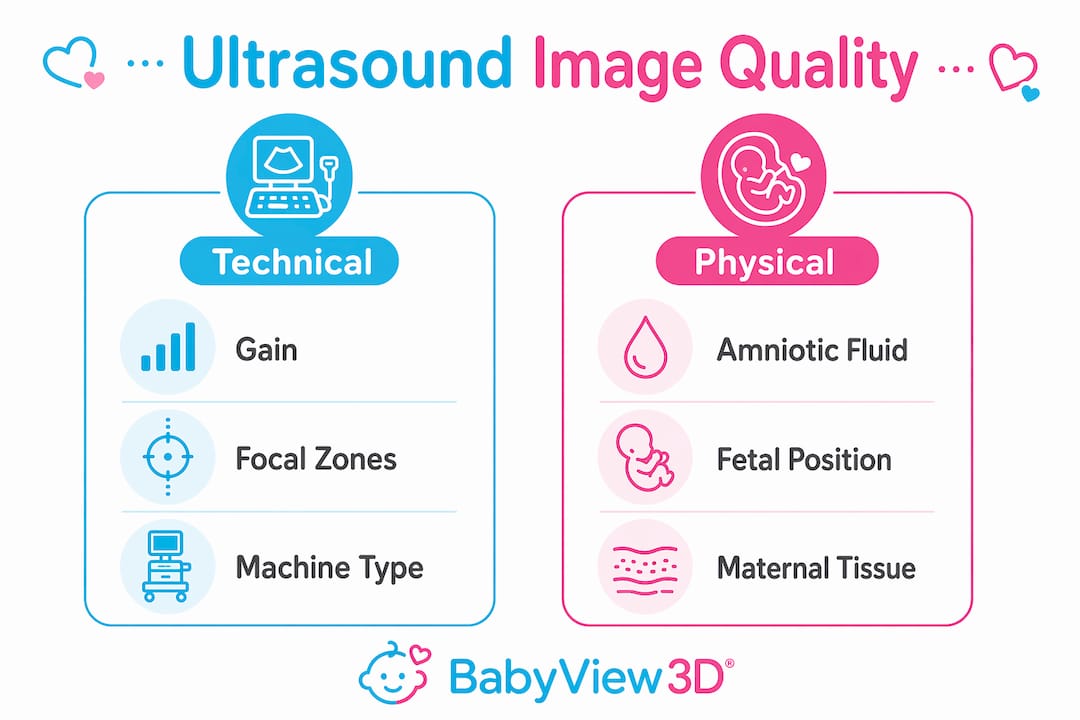
Bone and gas reflect nearly all ultrasound energy, creating shadows that block the view when a fetal face is pressed against the placenta or uterine wall. This is basic sound physics. When your baby tucks their chin to their chest or rests their face against the placenta, the acoustic path is blocked. No amount of gain adjustment fixes a shadow caused by bone or gas in the way.
Here are the main physical factors that shape what you see on screen:
- Fetal position. A baby facing the placenta or with limbs covering the face produces limited or obscured images regardless of machine quality. Repositioning through gentle movement or waiting is often the only solution.
- Amniotic fluid levels. Fluid acts as an acoustic window, transmitting sound waves cleanly to the baby. Insufficient amniotic fluid causes shadowing that no technical adjustment can fully overcome. More fluid around the face means cleaner, sharper images.
- Placental location. An anterior placenta sits between the probe and the baby, adding an extra layer of tissue the sound must pass through. This softens fine detail and increases the chance of shadowing.
- Body composition. A March 2026 study found that patient BMI has no relevant influence.html) on handheld ultrasound image quality compared to device familiarity and personal preference. This challenges a common assumption and shifts focus back to technique and positioning.
Sonographers consistently report that parents overestimate the impact of operator skill or equipment when anatomical factors are the actual limiting force. Understanding this prevents frustration and helps you ask better questions during your appointment.
Pro Tip: Drink 32 ounces of water in the two hours before your 3D or 4D scan. Better hydration supports amniotic fluid levels, which directly improves the acoustic window around your baby's face.
How do different ultrasound devices and innovations impact image quality?
Not all ultrasound machines produce the same results, and the gap between device types is wider than most parents realize. Handheld ultrasound devices have expanded access to prenatal imaging, but their output varies considerably depending on the specific model and the operator's familiarity with it.
| Device type | Image quality strengths | Key limitations |
|---|---|---|
| High-end clinical systems | Superior resolution, advanced 3D/4D rendering, HD Live color modes | Higher cost, clinic-based only |
| Mid-range portable systems | Good 2D clarity, adequate for standard prenatal checks | Limited 3D rendering depth and detail |
| Handheld ultrasound devices | Accessible, portable, useful for quick assessments | Quality varies by model; operator familiarity matters more than BMI |
The way professionals evaluate image quality is also changing. Traditional metrics like PSNR (Peak Signal-to-Noise Ratio) measure pixel-level differences but fail to capture how a human expert actually perceives image usefulness. AI-integrated evaluation frameworks have improved accuracy in predicting expert preferences for ultrasound image quality from 47.2% to 72.8%, using tools like the TinyUSFM-based framework. That jump represents a meaningful shift in how quality is measured and optimized across different systems.
For parents, this matters because it means the field is moving toward quality standards that reflect what actually looks good and useful to a trained eye, not just what scores well on a mathematical formula. Studios and clinics using prenatal ultrasound innovations aligned with these newer evaluation methods are better positioned to deliver images that genuinely satisfy families.
What practical tips help you get the best ultrasound images?
Knowing the science behind image quality gives you real leverage during your prenatal appointments. You are not a passive observer. You can take specific steps that meaningfully improve what you see.
- Choose the right gestational window. The 26 to 32 week range offers the best balance of fetal size, fat deposits under the skin, and available space in the uterus. Before 26 weeks, babies have less subcutaneous fat, which reduces surface detail. After 32 weeks, the uterus becomes crowded and repositioning is harder.
- Hydrate before your scan. Adequate fluid intake supports amniotic fluid levels, which function as the acoustic window between the probe and your baby. This is one of the few things you can directly control.
- Encourage fetal movement. Walking around the clinic hallway or drinking cold water before the scan can prompt your baby to shift position. A baby facing outward with clear fluid around the face produces dramatically better 3D images.
- Ask specific questions. Ask your sonographer about the probe frequency settings being used and whether the focal zone is positioned at the baby's face. These are not intrusive questions. They show you understand the process and help the sonographer focus on what matters to you.
- Understand 3D rendering modes. HD Live and similar rendering technologies add surface lighting and color to 3D images, making them look more lifelike. The underlying quality still depends on gain, focal zones, and fetal position. A well-rendered image of a poorly positioned baby will still disappoint.
- Manage expectations honestly. Some scans produce stunning images. Others produce limited ones despite perfect technique and optimal settings. Physical realities like fetal position or limited amniotic fluid create imaging limits that go beyond operator skill or machine quality. Accepting this makes the experience less stressful and more enjoyable.
For a deeper look at how sonographers optimize settings step by step, the 3D ultrasound process guide at Bbview3d's blog walks through each adjustment in plain language.
Key takeaways
Ultrasound image quality depends on the interaction of machine settings, sound physics, and fetal anatomy, and no single factor alone determines what you see on screen.
| Point | Details |
|---|---|
| Gain is the first fix | Reducing high gain is the most common and effective correction for grainy 3D images. |
| Focal zone placement matters | Misaligned focal zones cause blurring that persists even when all other settings are correct. |
| Anatomy often limits quality | Fetal position and amniotic fluid levels frequently control image clarity more than equipment does. |
| BMI is not the main variable | A 2026 study confirmed device familiarity and operator preference outweigh body composition in handheld scans. |
| AI is improving evaluation | New frameworks now predict expert image preferences with 72.8% accuracy, up from 47.2% with traditional metrics. |
Why I think parents deserve a more honest conversation about ultrasound images
Most families walk into a 3D or 4D scan expecting something close to a photograph. When the image comes out grainy or the baby's face is obscured, the immediate assumption is that something went wrong. Either the equipment was poor or the sonographer did not try hard enough. After working in prenatal imaging for years, I can tell you that assumption is almost always wrong.
The honest truth is that a baby pressing their face against the placenta at 30 weeks is a more powerful force than any machine setting. Sound physics does not negotiate. Bone blocks. Gas scatters. Fluid transmits. These are not failures of technology. They are the conditions of imaging a living, moving person inside another person.
What I find genuinely exciting is how AI-based evaluation tools are finally giving professionals a way to measure quality the way a human expert sees it, not just as a pixel ratio. That shift will eventually translate into better real-time feedback for sonographers and better images for families. We are not there yet, but the direction is clear.
My advice to every expectant parent: go in curious, not demanding. Ask questions. Understand that a blurry image of your baby's hand is still your baby's hand. And if the conditions are right, the image you get will be worth every minute of the wait.
— LENIER
See your baby in stunning detail with Bbview3d

Bbview3d has spent over 15 years helping families experience prenatal imaging at its best. Certified sonographers adjust gain, focal zones, and probe frequency in real time to get the clearest possible view of your baby, using HD Live technology across 3D, 4D, and 8K imaging formats. Every session is designed around your baby's position and your family's experience, not just a standard protocol. When conditions allow, the results are remarkable. Explore Bbview3d's prenatal imaging services to see available packages, locations across the United States, and current first-appointment offers. Your baby is worth seeing clearly.
FAQ
What is ultrasound image quality in prenatal scans?
Ultrasound image quality refers to the clarity, sharpness, and diagnostic usefulness of images produced during a prenatal scan. It is determined by machine settings like gain and focal zones, sound physics, and the baby's position in the womb.
Why does my 3D ultrasound look grainy?
Grainy 3D ultrasound images are most commonly caused by gain settings that are too high. The recommended fix is to reduce gain first, then increase smoothing, then adjust the probe angle.
Does body weight affect ultrasound image quality?
A March 2026 study found that patient BMI has no relevant influence on handheld ultrasound image quality. Device familiarity and operator preference are more significant factors than body composition.
What is the best time to get a 3D or 4D ultrasound?
The 26 to 32 week gestational window produces the best 3D and 4D images because the baby has developed subcutaneous fat for surface detail and there is still enough space and fluid for clear imaging.
Why is my baby's face hard to see in the ultrasound?
When a baby's face is pressed against the placenta or uterine wall, bone and gas reflect nearly all ultrasound energy, creating shadows that block the view. Gentle movement or waiting for the baby to reposition is the most effective solution.