
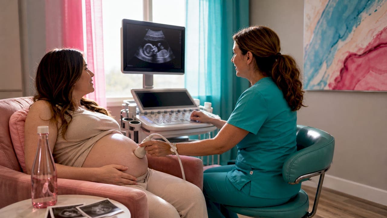
Ultrasound captures baby expressions by converting reflected sound waves into detailed 3D and 4D images that show your baby's face and movements in real time. This process, formally called sonographic surface rendering, gives expecting parents their first real look at a developing child's features before birth. The expressions you see, including yawns, smiles, and frowns, are not random. They reflect active neurological and muscular development happening inside the womb. Understanding why ultrasound captures baby expressions so clearly helps you appreciate both the technology and the biology behind every image.
Why ultrasound captures baby expressions: the technology explained
Ultrasound imaging works by sending high-frequency sound waves into the body. Those waves bounce off fetal tissues and return as echoes, which a computer processes into visual images. A standard 2D scan reads those echoes as flat, cross-sectional slices. That is enough to measure organs and check anatomy, but it does not show surface detail.

3D ultrasound assembles dozens of 2D image slices into a single still, three-dimensional surface view. The result looks like a photograph of your baby's face. 4D ultrasound adds the time dimension, turning that still image into live video so you can watch your baby move in real time. This is why 4D sessions feel so personal. You are not looking at a snapshot. You are watching your baby yawn or stretch as it happens.
The rendering software behind these images compiles 3D datasets from rapid 2D sweeps, then applies virtual lighting and angle selection to create the lifelike appearance of fetal faces. The result looks organic, but it is an algorithmic construction built from wave data. That distinction matters because it explains why image quality can vary so much from one session to the next.
- Sound waves transmit. The probe sends pulses into the uterus.
- Echoes return. Tissues at different depths reflect waves at different intensities.
- Software compiles. A computer stacks the echo data into a 3D model.
- Rendering applies. Virtual lighting creates the surface appearance you see on screen.
- 4D streams live. The process repeats fast enough to display real-time movement.
Pro Tip: Ask your sonographer to show you both a 3D still and a 4D live clip during your session. The live clip often captures the most expressive moments because babies move unpredictably.
When does ultrasound show the clearest baby face?
Timing is the single biggest factor in image quality. The optimal window for facial imaging is 26–32 weeks of gestation. During this period, your baby has built up enough subcutaneous fat to define cheekbones, lips, and a nose. The uterus still has enough space and amniotic fluid for the baby to shift positions, which gives the sonographer better angles.
Before 24 weeks, facial features are present but not yet filled out. The face can look skeletal or flat in surface renderings. After 34 weeks, the baby is larger and more cramped, making it harder to get a clear, unobstructed view of the face.
| Gestational window | Fat development | Fluid space | Expression visibility |
|---|---|---|---|
| Before 24 weeks | Minimal | Ample | Low |
| 24–26 weeks | Developing | Good | Moderate |
| 26–32 weeks | Well defined | Optimal | High |
| After 34 weeks | Fully developed | Reduced | Variable |
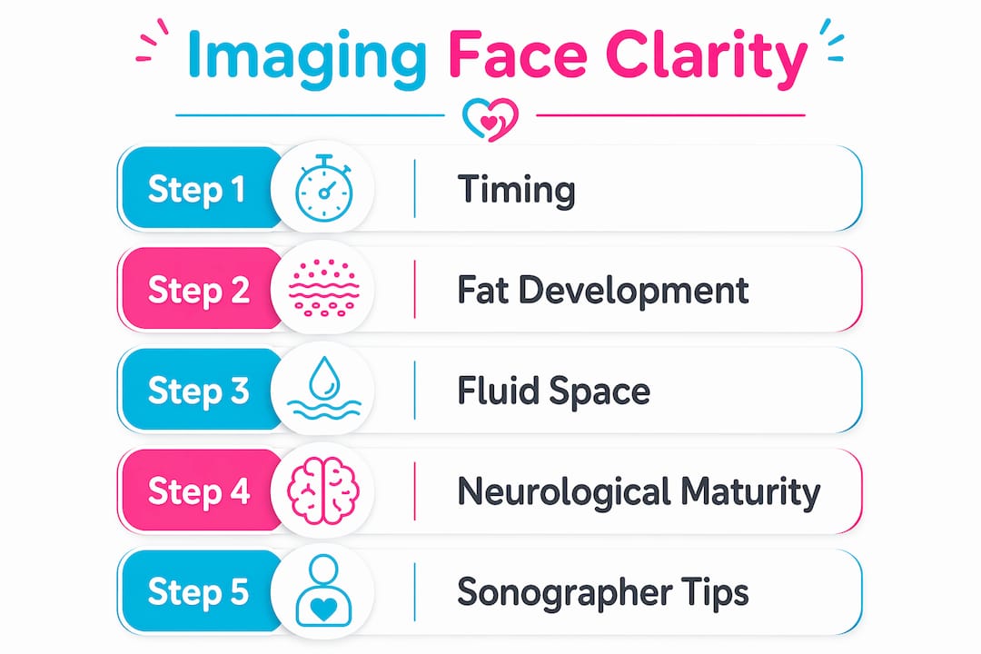
Fetal facial muscles and the neurological pathways that trigger them mature around this same 24–32 week window. That is not a coincidence. The biology and the imaging opportunity align. For parents who want to select the ideal timing for their session, scheduling between weeks 28 and 30 gives the best odds of a clear, expressive image.
What do baby facial expressions on ultrasound actually mean?
The expressions you see on a prenatal ultrasound are real muscle movements. They are not staged, and they are not random. Fetal facial expressions like frowning, yawning, and smiling begin appearing between 24 and 28 weeks as signs of neurological and muscular development. The brain is practicing. The muscles are rehearsing. Neither is communicating an emotion the way a newborn does.
True social smiles, the kind that respond to a face or a voice, only appear weeks after birth. What you see in the womb is the nervous system running drills. A yawn stretches the jaw muscles. A frown exercises the brow. A smile-like movement tests the orbicularis oris, the muscle that circles the mouth. Each one is a sign that development is on track.
This distinction matters because it helps you watch the images with the right frame of mind:
- Yawning signals jaw muscle activity and early respiratory practice.
- Frowning reflects brow muscle engagement tied to sensory input.
- Smiling movements exercise the lip and cheek muscles, not a social response.
- Eye squinting shows the orbicularis oculi muscle is developing.
- Tongue movements indicate oral motor development linked to feeding preparation.
"Fetal facial expressions captured by ultrasound are reflexive practice rather than social emotions. They provide reassuring evidence of neurological development." Source: Rosh MFM
Seeing your baby smile on screen is genuinely moving. Knowing it is a muscle rehearsal does not make it less meaningful. It makes it more remarkable. You are watching a nervous system build itself in real time.
What affects image quality and how sonographers improve it?
Even with advanced equipment, image quality is never guaranteed. Several physical factors can limit what the sonographer can capture. Baby position, amniotic fluid level, and placenta location all directly affect the clarity of 3D and 4D renderings.
A baby with both hands pressed against its face is one of the most common obstacles. The rendering software cannot cleanly separate hand data from face data, which produces distorted or merged features. A baby facing the placenta or uterine wall creates a similar problem. Clear amniotic fluid between the probe and the baby's face is the single most important physical condition for a sharp image. Even the most advanced software cannot compensate for a physical obstruction.
Operator skill is equally critical. A credentialed sonographer consistently produces better images than an inexperienced operator, even when both use the same equipment. Probe angle, pressure, and real-time adjustments to gain and depth settings all require trained judgment. Technology labels like HD Live or 5D describe software rendering modes, but the sonographer's hands determine whether the data going into that software is any good.
- Drink water in the hours before your appointment to support amniotic fluid clarity.
- Schedule during a time when your baby is typically active.
- Avoid a full meal right before the session, which can make the baby sluggish.
- If the baby's position is poor, a short walk or a cold drink can encourage movement.
Pro Tip: If your baby is not cooperating, ask your sonographer to pause for five minutes. A brief break often prompts a position change that opens up a clear facial view.
The emotional benefits of prenatal imaging are closely tied to image quality. A clear, detailed image creates a stronger sense of connection. That is why choosing a provider with certified sonographers and proper equipment matters as much as the technology itself.
Key Takeaways
Ultrasound captures baby expressions through 3D and 4D surface rendering of sound wave data, with the clearest results occurring between 26 and 32 weeks when fetal fat development and amniotic fluid levels are both optimal.
| Point | Details |
|---|---|
| Best imaging window | Schedule between 26 and 32 weeks for the clearest facial features and most expressive images. |
| Expressions are reflexive | Smiles, yawns, and frowns are neurological practice movements, not conscious emotions. |
| Sonographer skill matters | A certified sonographer's technique affects image quality more than the technology label alone. |
| Fluid and position are key | Clear amniotic fluid and an unobstructed baby face are the two biggest factors in image clarity. |
| 3D vs 4D purpose | 3D produces detailed still images; 4D adds live video so parents can watch expressions in real time. |
What seeing your baby's face actually does to you
Seeing a detailed image of your baby's face for the first time is one of those moments that does not need explanation. Parents who have been through it describe the same thing: the pregnancy becomes real in a way it was not before. That reaction is not sentimental. It is a known psychological effect of 3D and 4D ultrasound sessions, and it consistently deepens the emotional connection between parents and their developing child.
What I find most underappreciated is how much the science adds to the experience rather than taking away from it. Parents sometimes worry that knowing a smile is just a muscle exercise will make it feel less special. The opposite is true. When you understand that your baby's nervous system is actively building itself, and that the yawn you just watched is part of that process, the image carries more weight, not less.
The one expectation worth managing is the comparison between 3D and 4D sessions and the standard 2D anatomy scan. 3D and 4D scans are wonderful for bonding and seeing facial features, but they do not replace the 2D anatomy scan, which remains the standard for assessing internal organ development. These are two different tools with two different purposes. Treating them as interchangeable leads to disappointment in both directions.
My honest advice: go into a 3D or 4D session with the goal of connection, not diagnosis. Let the sonographer do their job. And if the baby's hand is covering its face for the first ten minutes, take a walk, drink some cold water, and try again. The moment you get a clear view of that face is worth the wait.
— LENIER
See your baby's face with Bbview3d
Bbview3d has spent over 15 years helping expecting parents see their baby's face in detail before birth. Certified sonographers use HD Live technology and advanced 3D and 4D imaging to capture the clearest possible views of your baby's expressions and movements.
Every session is designed around the emotional experience of connection, not just image capture. Bbview3d's centers across the United States offer comfortable environments where families can share the moment together. First-appointment discounts are currently available. Visit the Bbview3d services page to see available packages and book your session.
FAQ
Why do ultrasound captures baby expressions best at 26–32 weeks?
The 26–32 week window gives the baby enough subcutaneous fat to define facial features and enough amniotic fluid for clear imaging angles. Before 26 weeks, features are underdeveloped; after 34 weeks, space becomes too limited for good positioning.
Are the smiles and yawns I see on ultrasound real emotions?
No. Fetal facial expressions between 24 and 28 weeks are reflexive muscle movements driven by neurological development, not social emotions. True social smiles only appear several weeks after birth.
What is the difference between 3D and 4D ultrasound for seeing baby faces?
3D ultrasound produces a detailed still image of the baby's face, while 4D ultrasound adds live video so parents can watch facial expressions and movements as they happen in real time.
What can I do to get a clearer image of my baby's face?
Drink water in the hours before your appointment to support amniotic fluid levels, schedule during your baby's active period, and be prepared to take a short walk if the baby is not in a good position.
Does 3D or 4D ultrasound replace my standard prenatal scan?
No. 3D and 4D scans are supplemental tools for parental bonding and surface imaging. The standard 2D anatomy scan remains the medical standard for assessing internal organ development and fetal health.