
Ultrasound facial detail is the non-invasive imaging of facial structures beneath the skin using high-frequency sound waves, revealing muscles, bones, vessels, and tissue layers with precision. For expectant parents, this technology transforms a routine prenatal scan into a window onto your baby's developing face. Whether you are seeing the curve of a nose or the flutter of tiny lips, understanding how these images form helps you connect more deeply with what you are actually seeing. Bbview3d has spent over 15 years helping families experience this technology at its best, using 3D, 4D, and HD Live imaging to capture facial details that feel genuinely personal.
How ultrasound technology visualizes facial details beneath the skin
Ultrasound imaging works by sending high-frequency sound waves into tissue and recording the echoes that bounce back. Different tissue types, including bone, muscle, fat, and fluid, each reflect sound at different intensities. The scanner translates those differences into a grayscale or color-rendered image that maps the facial anatomy layer by layer. This is why a skilled sonographer can distinguish the outline of your baby's cheekbone from the soft tissue surrounding it.
The frequency matters enormously. Professional ultrasonic devices vibrate at approximately 28,000 cycles per second, generating a cavitation effect that interacts with tissue at a cellular level. Higher frequencies produce sharper, more detailed images of superficial structures, while lower frequencies penetrate deeper. Prenatal imaging typically balances both to capture a full picture of the developing face.
There are two fundamentally different categories of ultrasound technology that parents should understand:
- Diagnostic ultrasound creates images of anatomy safely and is the type used in every prenatal scan. It does not heat or alter tissue.
- Therapeutic ultrasound delivers energy intentionally to tissue for treatment purposes, such as skin tightening or collagen stimulation.
- HIFU (High-Intensity Focused Ultrasound) targets deep tissue layers for cosmetic or clinical outcomes and is never used in prenatal imaging.
- Handheld diagnostic scanners like the Clarius L20 provide real-time, high-resolution imaging that practitioners use to map facial anatomy during procedures.
Diagnostic ultrasound is non-invasive and safe for fetal imaging, which is the critical distinction for expectant parents. Therapeutic devices like Ultherapy or HIFU systems are entirely separate tools designed for adults seeking skin rejuvenation. Knowing this difference removes any confusion about safety.
| Technology | Primary use | Imaging capability |
|---|---|---|
| Diagnostic ultrasound | Fetal and anatomical imaging | Full real-time visualization |
| HIFU systems | Skin tightening, collagen remodeling | Limited or no live imaging |
| Ultherapy PRIME | Facial rejuvenation in adults | Real-time targeting of SMAS layer |
| Clarius L20 scanner | Clinical facial anatomy mapping | Split-screen, real-time display |
Pro Tip: Ask your sonographer to explain what you are seeing in real time during the scan. Practitioners using HD Live technology can narrate each facial feature as it appears on screen, turning the session into an educational experience rather than a passive one.
What are the benefits of ultrasound facial imaging for families?
The most direct benefit of detailed prenatal facial imaging is emotional. Seeing your baby's face, even in utero, triggers a bonding response that parents consistently describe as transformative. Studies on prenatal attachment confirm that visual contact with fetal features accelerates parental connection before birth. This is not a minor side effect of the technology. It is one of its most significant outcomes.
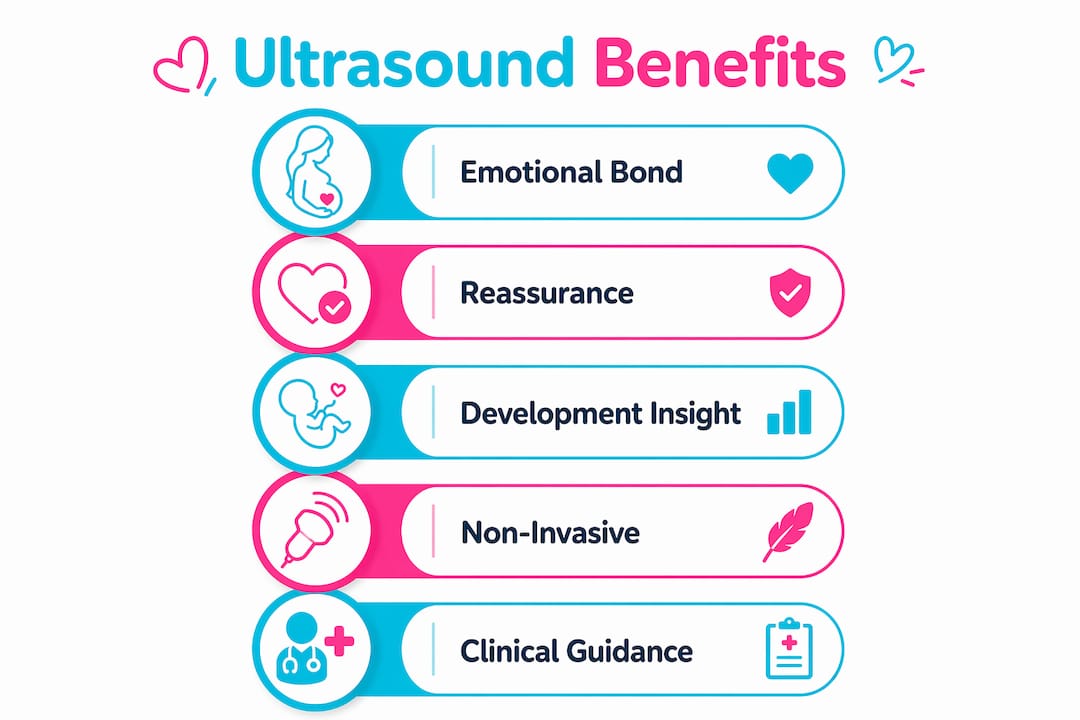
Beyond bonding, detailed facial imaging provides reassurance about fetal development. Parents can observe facial symmetry, the formation of the palate, and the movement of facial muscles during expressions like yawning or sucking. These observations give families confidence that development is progressing normally, which reduces prenatal anxiety in a meaningful way.
The benefits of ultrasound facials in clinical contexts, including non-invasive delivery and longer-lasting effects compared to traditional methods, parallel the advantages of diagnostic prenatal imaging. Both rely on the same core principle: precise, gentle interaction with tissue that produces results without physical trauma.
Advanced imaging formats like 3D and HD Live have significantly raised the standard for what families can see. Where older 2D scans showed flat cross-sections, HD Live technology renders light and shadow across the baby's face, producing images that look almost photographic. The role of HD Live technology in prenatal scans is specifically to improve the clarity of soft tissue surfaces, which is exactly where facial features live.
Key advantages for expectant families include:
- Visualization of facial expressions, including smiling, yawning, and eye movements
- Confirmation of structural development including lip formation and nasal bone presence
- Creation of keepsake images and videos that families can share and preserve
- Reduced anxiety through direct visual evidence of healthy fetal activity
- Early identification of features that may warrant follow-up with an OB or specialist
How ultrasound facial detail aids clinical procedures safely
Outside of prenatal care, explaining ultrasound facial detail becomes critical in aesthetic medicine. Practitioners performing facial injections, filler placements, or dissolving procedures now use real-time ultrasound imaging to map what lies beneath the skin before touching a needle to a patient's face. This is not a luxury. It is rapidly becoming the standard of care.
Real-time ultrasound mapping allows practitioners to visualize facial vascular anatomy and previously placed fillers, directly reducing complications during procedures. The implication is significant: a practitioner who can see a blood vessel before injecting near it is far less likely to cause a vascular occlusion, one of the most serious risks in facial aesthetics.
The Clarius L20 scanner is a specific example of how portable technology has changed clinical practice. Scans can identify target structures in as little as 20 seconds, giving practitioners a rapid anatomical map before any procedure begins. This speed makes ultrasound guidance practical in a busy clinical setting, not just in research environments.
Here is how a typical ultrasound-guided facial procedure unfolds:
- The practitioner applies gel to the treatment area and places the transducer against the skin.
- A real-time image appears on screen, showing tissue layers, vessels, and any existing filler deposits.
- The practitioner identifies safe injection zones and marks them based on the live image.
- The procedure proceeds with the ultrasound running, allowing continuous monitoring.
- Post-procedure imaging confirms correct placement and checks for any vascular involvement.
"Ultrasound allows me to see what I cannot feel. The difference between guessing and knowing is the difference between a safe outcome and a complication." This perspective, shared widely among dermatologists who use ultrasound guidance, reflects a shift in how the field approaches patient safety.
Ultherapy and HIFU devices differ primarily in their real-time imaging capability, which directly affects precision in skin tightening treatments. Ultherapy's built-in imaging allows practitioners to target the SMAS layer specifically, the same fibromuscular layer addressed in surgical facelifts. This level of precision is only possible because the device shows the practitioner exactly where the energy is being delivered.
Pro Tip: If you are considering any facial aesthetic procedure, ask your provider whether they use ultrasound guidance. Practitioners who use tools like the Clarius L20 or Ultherapy PRIME are working with a measurably higher safety standard than those operating without imaging.
What to expect during a prenatal ultrasound facial imaging session
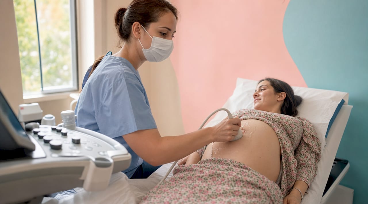
A prenatal ultrasound session focused on facial detail typically runs between 20 and 45 minutes, depending on the package and the baby's position. The experience is comfortable and requires no special preparation beyond arriving with a moderately full bladder, which improves image clarity in early sessions. After the second trimester, hydration matters more than bladder fullness.
Here is what happens during a typical session at a facility like Bbview3d:
- A certified sonographer applies warm gel to the abdomen and moves the transducer to locate the baby's face.
- The practitioner adjusts the imaging angle to capture the face in profile and then in a forward-facing view.
- HD Live or 3D rendering is applied in real time, adding depth and shadow to the facial image.
- Parents watch on a large monitor as features like the nose, lips, cheeks, and eyes become visible.
- The session is recorded, and families receive digital images or video keepsakes to take home.
Understanding the visual feedback makes the experience far richer. A split-screen display, common in advanced scanning suites, shows both the raw ultrasound data and the rendered 3D image simultaneously. This helps parents connect the technical scan with the recognizable face they are seeing. Learning to read ultrasound image enhancement before your session gives you a head start on interpreting what appears on screen.
The baby's position significantly affects image quality. Babies facing forward with their face near the uterine wall produce the clearest facial detail. If the baby is turned away, sonographers use gentle repositioning techniques or ask the parent to walk briefly to encourage movement. Patience during this part of the session almost always results in a clear, detailed image.
Key takeaways
Ultrasound facial detail imaging works because high-frequency sound waves reflect differently off bone, muscle, and soft tissue, producing precise layered images that reveal facial anatomy in real time.
| Point | Details |
|---|---|
| Diagnostic vs. therapeutic ultrasound | Diagnostic ultrasound is safe for fetal imaging; therapeutic types like HIFU are for adult skin treatments only. |
| HD Live improves facial clarity | 3D and HD Live rendering adds light and shadow to fetal face images, producing near-photographic detail. |
| Real-time imaging raises safety | Tools like the Clarius L20 identify facial structures in 20 seconds, reducing procedural risks significantly. |
| Bonding starts with seeing | Visual contact with fetal facial features during scans accelerates parental attachment before birth. |
| Session preparation matters | Baby position and maternal hydration directly affect the quality of facial detail captured during a scan. |
What 15 years of watching families see their baby's face has taught me
The moment a parent recognizes their baby's nose or catches a yawn on screen is not a small thing. I have watched it happen hundreds of times, and it never becomes routine. What strikes me most is how much the quality of the image changes the quality of that moment. A blurry, flat 2D scan produces recognition. A high-resolution HD Live image produces something closer to meeting.
What most articles on this topic miss is the cognitive shift that detailed imaging creates. When parents can see the facial muscles moving, the lips pressing together, the eyes tracking beneath closed lids, they stop thinking of the pregnancy in abstract terms. The baby becomes a specific person with a specific face. That shift has real effects on prenatal behavior, on stress levels, and on how prepared parents feel for birth.
I am also convinced that the clinical and prenatal uses of ultrasound facial detail are more connected than most people realize. The same technology that helps a dermatologist avoid a blood vessel during a filler injection is the technology that lets a sonographer show you the exact curve of your baby's upper lip. Precision imaging serves both safety and connection. That is a rare combination worth understanding deeply.
The step-by-step 3D ultrasound process is worth reviewing before your first session. Families who arrive knowing what to expect engage more actively, ask better questions, and leave with a richer experience. The technology does the heavy lifting, but your attention during the session determines how much you take away from it.
— LENIER
See your baby's face in extraordinary detail with Bbview3d
Bbview3d uses 3D, 4D, and 8K HD Live ultrasound technology to capture your baby's facial features with a level of clarity that standard prenatal scans simply cannot match. With over 15 years of experience and certified sonographers at multiple U.S. locations, every session is designed to give your family a genuine connection to your developing baby. Explore ultrasound imaging services built specifically for expectant families, from first-look packages to full keepsake sessions with digital images and video. Browse the Bbview3d gallery to see the level of facial detail that families across the country have experienced, and book your first appointment with a limited-time discount for new clients.
FAQ
What is ultrasound facial detail in prenatal imaging?
Ultrasound facial detail refers to the high-resolution visualization of a baby's facial anatomy, including muscles, bones, and soft tissue, using reflected sound waves. Advanced formats like 3D and HD Live rendering make individual features like the nose, lips, and cheeks clearly recognizable.
Is ultrasound safe for imaging my baby's face?
Diagnostic ultrasound is non-invasive and safe for fetal imaging, which is the type used in all prenatal scans. It does not deliver heat or energy to tissue the way therapeutic devices like HIFU do.
How does HD Live technology improve facial image quality?
HD Live applies light-source rendering to 3D ultrasound data, adding depth and shadow to the baby's face in real time. The result is a near-photographic image that shows surface texture and facial expressions far more clearly than standard 2D scans.
What affects the quality of facial detail in a prenatal scan?
Baby position is the single largest factor. Babies facing forward with their face near the uterine wall produce the clearest images. Maternal hydration and gestational age, with the optimal window typically between 26 and 32 weeks, also affect image sharpness significantly.
How is prenatal ultrasound different from ultrasound facial treatments for adults?
Prenatal ultrasound uses diagnostic technology to image anatomy without altering tissue. Adult ultrasound facial treatments, including HIFU skin tightening and therapeutic devices, deliver focused energy to stimulate collagen or cleanse pores. The two categories use different frequencies, intensities, and goals entirely.