
A guide to creating ultrasound milestones is a structured plan for tracking and celebrating each major fetal imaging appointment throughout pregnancy. These milestones cover the early viability scan at 6–8 weeks, the nuchal translucency (NT) scan at 11–14 weeks, and the detailed anatomy scan at 18–22 weeks. Each appointment delivers both clinical data and a memory worth keeping. Modern tools, from 3D/4D imaging technology to digital photo albums, make it easier than ever to document your baby's development with ultrasound and build a record you will treasure for life.
What are the key ultrasound milestones during pregnancy?
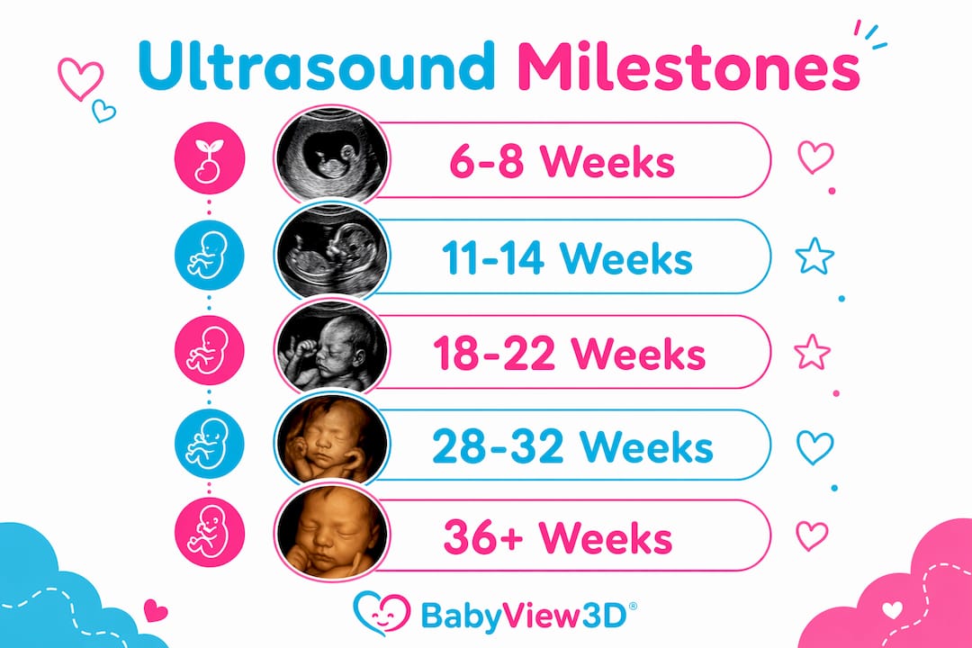
Ultrasound milestones are the scheduled imaging appointments that mark distinct stages of fetal growth. Each scan has a specific clinical purpose, and knowing what to expect at each one helps you prepare emotionally and practically.
The first trimester scans
The first ultrasound at 6–8 weeks confirms that the pregnancy is viable and establishes your due date using Crown-Rump Length (CRL), a measurement accurate to within 3–8 days. This is often the scan where parents hear the heartbeat for the first time. That moment alone makes it one of the most emotionally significant appointments in the entire pregnancy.
The NT scan follows at 11–14 weeks and screens for chromosomal abnormalities by measuring the fluid at the back of the baby's neck. It is typically combined with a blood test for the most accurate risk assessment. Many parents also get their first clear profile image of the baby's face at this appointment.
The second trimester anatomy scan
The anatomy scan at 18–22 weeks is the most detailed imaging appointment in a standard pregnancy. Sonographers assess fetal organs, limbs, the placenta, and amniotic fluid levels. Common measurements include biparietal diameter (BPD), head circumference (HC), abdominal circumference (AC), and femur length (FL). Gender identification is optional at this stage, making it a natural moment for a gender reveal if that is part of your plan.
Third trimester growth scans
Late pregnancy scans focus on fetal positioning, growth rate, and placental health. These are especially common for higher-risk pregnancies or when earlier scans flagged anything worth monitoring. They are less about dramatic reveals and more about confirming that your baby is on track for a healthy delivery.
| Scan | Timing | Primary Purpose |
|---|---|---|
| Early viability scan | 6–8 weeks | Confirm pregnancy, establish due date via CRL |
| Nuchal translucency scan | 11–14 weeks | Screen for chromosomal risk |
| Anatomy scan | 18–22 weeks | Full organ and structural assessment |
| Growth and positioning scan | 28–36 weeks | Monitor growth, check baby's position |
Pro Tip: Ask your provider at the anatomy scan whether the baby's position allows for a clear face image. If not, a follow-up scan is sometimes scheduled, which gives you a second chance at that keepsake shot.
How to set ultrasound goals and build your milestone checklist
Setting goals before each appointment transforms a routine medical visit into a meaningful milestone. The first step is separating medically indicated scans from elective keepsake sessions.
Keepsake or gender-reveal-only scans do not qualify as billable medical necessity under current CPT guidelines. That distinction matters because it affects cost, insurance coverage, and how you schedule them. Medical scans are ordered by your OB or midwife. Elective 3D/4D sessions are booked separately, often at specialized imaging studios.
Building your ultrasound milestone checklist
Use this framework to organize your ultrasound development timeline from the first trimester through delivery:
- Confirm your medically scheduled scans. Ask your provider which scans are standard for your pregnancy and whether any additional monitoring is recommended.
- Identify your personal milestone goals. These might include capturing the first heartbeat, getting a clear 3D face image, or timing a gender reveal.
- Schedule elective sessions strategically. The 26–30 week window often produces the clearest 3D/4D images because the baby has enough fat under the skin to show facial features clearly.
- Prepare questions for each appointment. Write them down beforehand so you do not forget in the moment.
- Plan your documentation method. Decide whether you will use a physical scrapbook, a digital album app, or a dedicated pregnancy journal.
Pro Tip: Tell your sonographer at the start of the session what images matter most to you. A good technician will prioritize those shots while still completing the clinical checklist.
When you understand your ultrasound report, focus on the summary or conclusion section rather than the raw biometric measurements. Those numbers are written for clinical interpretation. The summary tells you what actually matters in plain language.
How to document and celebrate each ultrasound milestone
Documenting your baby's development with ultrasound is about more than saving images. It is about creating a record that tells the story of your pregnancy from the first flicker of a heartbeat to the final growth check before birth.
Preparing for each appointment
Arrive with a list of questions, a full bladder if required, and a charged phone or camera for any images you are allowed to photograph from the screen. Bring your partner or a support person when possible. Shared experiences anchor memories more deeply than solo ones.
Capturing and storing your images
Most clinics provide printed images or digital files after each scan. Store these in a dedicated folder immediately after each appointment so they do not get lost in your camera roll. Apps like Pregnancy+ and The Bump offer built-in milestone trackers where you can attach ultrasound images to specific weeks.
"Every image, even a blurry one, is a timestamp. It says: your baby existed, was growing, and was loved, right at that moment."
For families who want higher-quality images, advanced ultrasound features like HD Live and 8K resolution capture details that standard 2D scans simply cannot match. The difference between a flat gray silhouette and a lifelike 3D face image is significant when you are building a keepsake collection.
Celebrating your milestones
- Create a milestone card for each scan with the date, gestational week, and one detail you learned or saw.
- Use the anatomy scan image as the centerpiece of a gender reveal if that fits your plan.
- Build a scrapbook or digital photo book that grows with each appointment.
- Share milestone images with family through a private social media group or a shared photo album.
Handling difficult news requires a different kind of preparation. If a scan requires follow-up, write down exactly what the provider said before you leave the office. Emotions can blur details quickly. A clear written record helps you ask better questions at the next appointment.
What are the most common ultrasound challenges and how do you handle them?
Incomplete scans are more common than most parents expect. A review of over 15,000 prenatal exams found significant variation in scan completeness, with higher rates of incomplete anatomy scans in patients with higher maternal BMI and in scans performed before 19 weeks. An incomplete scan is not a sign that something is wrong. It is a normal part of clinical practice.
The most common reason for an incomplete scan is baby position. If the baby is facing the wrong way or has a limb blocking the view, the sonographer cannot capture every required image in one session. This leads to a follow-up scan, which is actually an opportunity to get more images and spend more time with your baby on screen.
Expert sonographers use a defined pivot plan to keep sessions productive when the baby is uncooperative. This includes switching between capturing the profile, hands, feet, and baby stretching to maintain session flow and keep families engaged. Scripted micro goals, like holding a clear profile image for five seconds, help build consistency even in challenging sessions.
"A good sonographer does not panic when the baby moves. They pivot, stay calm, and keep narrating. That calm is contagious."
Pro Tip: If you have an elective 3D/4D session and the baby is not cooperating, drink a small amount of cold juice 20–30 minutes before the appointment. The sugar and temperature change often increase fetal movement and repositioning.
Families find ultrasound milestones most rewarding when sonographers engage with clear narration and calm pacing throughout the session. If your technician is not narrating, ask them to explain what they are seeing. You are allowed to ask questions during the scan.
Key takeaways
Documenting your ultrasound development timeline with clear goals and a structured checklist turns routine medical appointments into lasting family memories.
| Point | Details |
|---|---|
| Know your scan timeline | The four core milestones are at 6–8, 11–14, 18–22, and 28–36 weeks, each with a distinct purpose. |
| Separate medical from elective scans | Medically indicated scans are covered differently than keepsake sessions; plan and budget for both. |
| Build a milestone checklist | Define personal goals like first heartbeat capture or 3D face image before each appointment. |
| Document immediately after each scan | Store images in a dedicated folder or app right after each appointment to avoid losing them. |
| Incomplete scans are normal | Follow-up scans due to baby position or timing are routine, not a cause for alarm. |
Why milestone planning changed how i experience pregnancy scans
I have sat in on hundreds of prenatal ultrasound sessions over the years, and the families who get the most out of them are never the ones who simply show up and wait. They are the ones who walked in knowing what they wanted to see and what questions they needed answered.
Most parents treat ultrasound appointments as passive events. You lie down, the technician scans, you get a photo, and you leave. That approach misses most of what these appointments can offer. When you treat each scan as a milestone with a specific goal, the entire experience changes. You are present. You are asking questions. You are building a story.
The clinical side of ultrasound planning matters, but it is not the whole picture. I have seen parents leave an anatomy scan feeling anxious because they fixated on measurements they did not understand. I have also seen parents leave the same type of scan feeling connected and confident because they focused on the summary, asked about what they saw, and left with a clear image of their baby's face. The difference was preparation, not the scan itself.
One thing most guides skip: the emotional recovery after a difficult scan. If a result requires follow-up, the worst thing you can do is go home and search symptoms online. Write down what the provider said, call a trusted friend or family member, and wait for the follow-up appointment before drawing conclusions. Anxiety fills gaps with worst-case scenarios. A structured milestone plan gives you something concrete to focus on instead.
Cherish every image, even the blurry ones. A perfect photo is not the point. The point is that you were there, paying attention, and building a connection with your baby before they were born. That is what ultrasound milestones are really for.
— LENIER
See your baby's milestones in stunning detail with Bbview3d
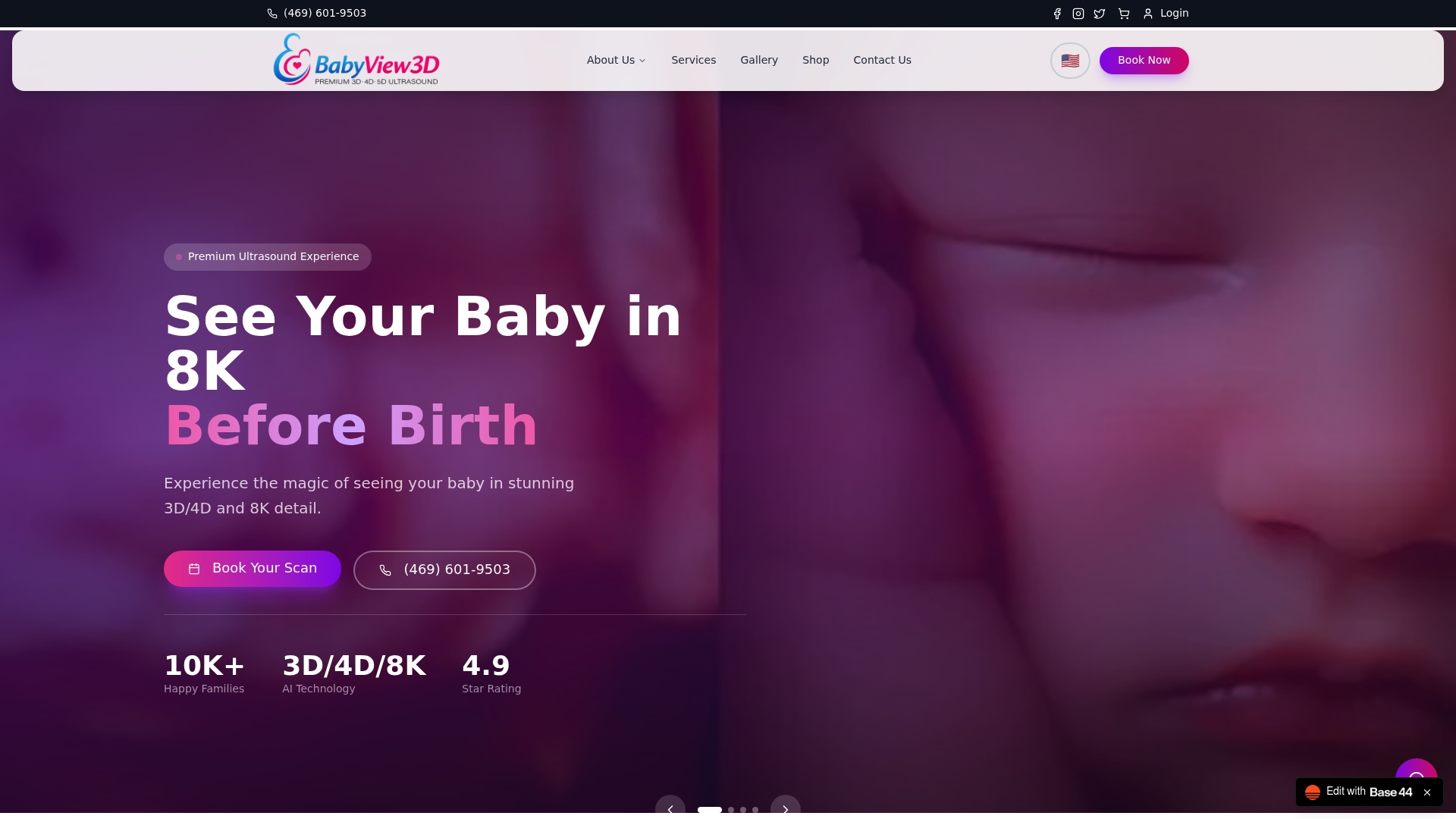
Bbview3d specializes in prenatal ultrasound experiences that go far beyond a standard clinic scan. With over 15 years of experience and certified sonographers at multiple locations across the United States, Bbview3d offers 3D, 4D, and 8K HD Live imaging sessions designed to capture the moments that matter most. Whether you are planning a gender reveal, a first face image, or a complete keepsake package, their ultrasound imaging services include visual summaries, digital galleries, and take-home keepsakes that bring your milestone checklist to life. First-time visitors can access a limited introductory offer. Browse their keepsake store to find the right package for your family.
FAQ
When does the first ultrasound milestone happen?
The first ultrasound occurs at 6–8 weeks and confirms pregnancy viability using Crown-Rump Length to date the pregnancy within 3–8 days. This is typically when parents hear the heartbeat for the first time.
How do i document baby's development with ultrasound?
Store images in a dedicated folder or pregnancy app immediately after each scan, and attach a note with the gestational week and key findings. Apps like Pregnancy+ and The Bump include built-in milestone trackers for this purpose.
What is the difference between a medical and elective ultrasound?
Medical ultrasounds are ordered by your provider and have a clinical indication. Elective keepsake scans do not qualify as billable medical necessity and are scheduled separately at your own cost.
Why was my anatomy scan incomplete?
Incomplete anatomy scans are common and are most often caused by baby position or early timing before 19 weeks. A follow-up scan is standard practice and does not indicate a problem with your pregnancy.
When is the best time for a 3d or 4d keepsake session?
The 26–30 week window produces the clearest 3D and 4D images because the baby has developed enough subcutaneous fat to show defined facial features. For ideal ultrasound timing, schedule your elective session within this window for the best results.