

3D ultrasound is defined as a prenatal imaging technique that uses high-frequency sound waves to reconstruct detailed surface images of the fetus in real time, while fetal MRI uses magnetic fields to capture cross-sectional views of internal structures. For most expectant parents, the 3D ultrasound vs MRI comparison comes down to one practical truth: ultrasound is your first-line tool, and MRI steps in when deeper diagnostic clarity is needed. Both are radiation-free. Both serve distinct roles. Understanding which does what will help you ask the right questions at every prenatal appointment.
1. What are the technical differences between 3D ultrasound and MRI?
The core difference is how each method captures images. 3D ultrasound uses sound waves to reconstruct surface images rapidly and portably, while MRI uses magnetic fields to capture detailed cross-sectional images but requires a specialized suite and longer scan time. A standard ultrasound session takes minutes. An MRI exam takes tens of minutes and demands that the patient remain still throughout.
Portability is another major gap. Ultrasound machines travel to clinics, hospitals, and even mobile units. MRI scanners are fixed installations that require dedicated rooms with shielding. That difference alone explains why ultrasound handles routine prenatal care while MRI is reserved for complex cases.
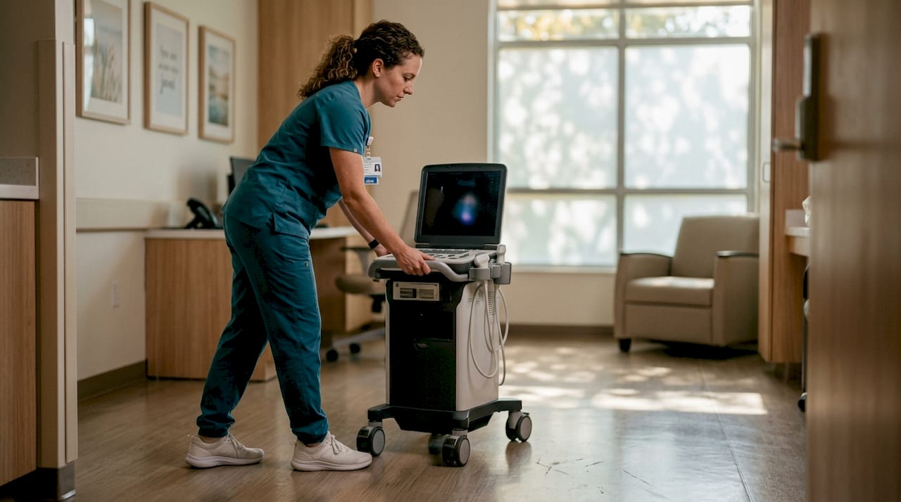
Safety profiles are similar in one key way: neither modality uses ionizing radiation. MRI does require screening for metallic implants and medical devices, which adds a preparation step that ultrasound does not. MRI requires specialized suites and is often more costly, making it less accessible for routine visits.
Operator skill also plays a larger role in ultrasound than in MRI. MRI acquisition follows standardized protocols. Ultrasound image quality depends heavily on the sonographer's technique, probe angle, and real-time adjustments. Certified sonographers with volumetric imaging experience consistently produce clearer 3D results.
Pro Tip: Ask your provider whether their sonographer holds specific certification in 3D volumetric imaging. Operator experience directly affects the detail you see in your images.
| Feature | 3D Ultrasound | Fetal MRI |
|---|---|---|
| Technology | High-frequency sound waves | Magnetic fields and radiofrequency |
| Scan duration | Minutes | Tens of minutes |
| Portability | Yes | No |
| Radiation | None | None |
| Cost | Lower | Higher |
| Primary use | Surface and structural visualization | Deep internal tissue contrast |
2. When is the best timing for 3D ultrasound during pregnancy?
Timing determines everything with 3D imaging. The optimal gestational window for high-quality 3D ultrasound imaging is between 24 and 32 weeks, when amniotic fluid volume is adequate and the fetus has developed enough facial definition to produce clear images. Before 24 weeks, the fetus lacks the fat deposits that give surface images their detail. After 32 weeks, the fetus descends into the pelvis and fluid decreases, both of which reduce image clarity.
Within that window, there are two distinct purposes for a 3D scan. Diagnostic scans, ordered by your OB or maternal-fetal medicine specialist, assess structural anomalies and require a certified sonographer. Elective bonding scans serve visualization and emotional connection purposes, giving parents a detailed look at facial features and movements in real time. Both types use the same technology, but their clinical weight differs.
What 3D ultrasound shows best includes facial features, limb surfaces, and external structural details. It does not show internal organ detail with the same clarity as MRI. Parents who expect to see brain or spine anatomy in a 3D scan will be disappointed. That level of internal detail requires a different tool entirely.
- Scan between 24 and 32 weeks for the clearest surface images
- Hydrate well before your appointment to support amniotic fluid volume
- Schedule in the morning when fetal activity tends to be higher
- Understand whether your scan is diagnostic or elective before you book
Pro Tip: Drink 32 ounces of water two hours before your 3D ultrasound. Better hydration supports amniotic fluid clarity, which directly affects image quality.
3. What advantages does MRI offer over 3D ultrasound in prenatal imaging?
MRI's defining strength is soft tissue contrast. MRI offers superior soft tissue contrast and multiplanar imaging, making it the preferred tool for evaluating complex fetal abnormalities such as brain and spine anomalies when ultrasound findings are inconclusive. No ultrasound technique, including 3D, matches MRI for internal organ visualization.
The clinical pathway is clear: ultrasound comes first, and MRI follows when a finding needs further investigation. A maternal-fetal medicine specialist typically orders fetal MRI after a detailed anatomy scan raises a question that ultrasound cannot resolve. This staged approach reflects both safety and cost efficiency.
MRI does carry practical drawbacks for pregnant patients:
- Longer scan times require extended stillness, which is physically demanding in the third trimester
- Patients must be screened for metallic implants, pacemakers, and other devices before entering the scanner
- MRI exams are longer and require preparation, including metal screening, making them less accessible and comfortable than ultrasounds
- Cost is significantly higher, and not all facilities offer fetal MRI
- Availability is limited to larger medical centers in many regions
The discomfort factor is real. Lying still inside a narrow MRI bore for 30 to 45 minutes while pregnant is challenging. Most facilities offer open-bore or wide-bore MRI options for patient comfort, but these are not universally available.
4. How do 3D ultrasound and MRI compare in clinical outcomes?
Clinical evidence places these two tools in very different categories. 3D ultrasound improves visualization of facial and surface anomalies but has not consistently shown improved clinical outcomes over 2D ultrasound in most pregnancies. One study found that only 4.8% of cases showed changed clinical care due to 3D ultrasound findings. That number tells you something important: 3D ultrasound adds visual detail, but it rarely changes what your doctor decides to do next.
MRI, by contrast, is ordered precisely because it is expected to change clinical management. When a 2D anatomy scan flags a potential brain abnormality, fetal MRI can confirm, rule out, or better characterize the finding. That information directly affects counseling, delivery planning, and postnatal care.
The diagnostic pathway prioritizes ultrasound first and MRI only when further investigation is needed, reflecting a staged, safe approach to prenatal imaging. This is not a hierarchy of quality. It is a logical sequence based on what each tool does best.
Standard 2D ultrasound remains the backbone of prenatal care. Both 3D ultrasound and MRI are supplements to it, not replacements. Parents who understand this distinction make better decisions about which scans to pursue and why.
| Imaging type | Primary clinical role | Changes management? |
|---|---|---|
| 2D ultrasound | Routine anatomy and growth monitoring | Yes, frequently |
| 3D ultrasound | Surface detail and visualization | Rarely (4.8% of cases) |
| Fetal MRI | Complex internal anatomy clarification | Yes, when ordered |
5. Which prenatal imaging option should expectant parents choose?
The answer depends on your clinical situation. 3D ultrasound is the right first choice for most expectant parents. It is affordable, widely available, and gives you detailed surface images of your baby's face and body. 3D ultrasound excels at real-time surface visualization while MRI is valued for detailed internal tissue contrast, making them complementary tools rather than competing ones.
MRI belongs in the picture only when your care team identifies a finding that ultrasound cannot fully characterize. High-risk pregnancies, suspected brain or spinal cord anomalies, and complex organ malformations are the situations where fetal MRI earns its place. You do not need to request an MRI. Your provider will recommend it if the clinical picture calls for it.
Practical factors that affect your choice include:
- Timing: Book your 3D ultrasound between 24 and 32 weeks for the best results. Learn more about ideal ultrasound timing to plan your session effectively.
- Fetal position: A baby facing away from the probe will produce poor surface images. Flexibility in rescheduling matters.
- Image quality factors: Image quality depends on fetal position and fluid volume, not just machine quality, often requiring flexibility in scan timing.
- Budget: 3D ultrasound sessions are significantly less expensive than fetal MRI and are often available without a physician referral for elective purposes.
- Accessibility: Fetal MRI requires a specialized center. 3D ultrasound is available at most OB practices and dedicated imaging studios.
Pro Tip: If your baby is in a poor position during your 3D scan, ask the sonographer to pause and have you walk around for 10 minutes. Movement often shifts fetal position enough to get a clear image.
6. What role does the ACOG guidance play in prenatal imaging decisions?
Professional guidelines shape how providers use these tools. The American Congress of Obstetricians and Gynecologists (ACOG) and the American Institute of Ultrasound in Medicine recommend that 3D and 4D ultrasounds serve as adjuncts to standard 2D scans. They do not recommend 3D ultrasound for routine use, and they discourage non-medical keepsake imaging performed outside a clinical setting.
This guidance does not mean elective 3D scans are harmful. It means they are not medically necessary for most pregnancies. Elective 3D and 4D scans can show facial expressions and movements in real time, enhancing the parent experience and strengthening emotional connection before birth. That value is real, even if it is not clinical.
The distinction between a diagnostic scan and a bonding scan matters for how you interpret what you see. A bonding scan at a professional studio gives you beautiful images and a memorable experience. A diagnostic scan at a maternal-fetal medicine practice gives you clinical information. Both have their place. Neither replaces the other.
Understanding ultrasound image quality factors before your appointment helps you set realistic expectations for what your images will look like.
Key Takeaways
3D ultrasound is the practical, accessible choice for prenatal visualization, while fetal MRI is a specialized diagnostic tool reserved for complex cases that ultrasound cannot fully resolve.
| Point | Details |
|---|---|
| 3D ultrasound is first-line imaging | It is affordable, portable, and ideal for surface visualization between 24 and 32 weeks. |
| MRI serves complex cases | Fetal MRI is ordered after ultrasound raises unresolved questions about internal anatomy. |
| Clinical impact of 3D is limited | Only 4.8% of cases showed changed clinical management from 3D ultrasound findings. |
| Timing and position affect quality | Image clarity depends on fetal position and amniotic fluid, not just equipment. |
| Both tools are complementary | 2D ultrasound remains the baseline; 3D and MRI each add specific value on top of it. |
What I've learned from watching parents navigate prenatal imaging
Most expectant parents walk into their first 3D ultrasound appointment expecting a medical verdict. They leave with beautiful images and sometimes a lingering question: "But what does it mean?" That confusion is worth addressing directly.
3D ultrasound is not a diagnostic replacement for your anatomy scan. It is a visualization tool, and a genuinely moving one. Seeing your baby's face in detail before birth creates a connection that flat 2D images simply cannot match. That emotional value is legitimate. You do not need to justify wanting it.
MRI, on the other hand, is not something to fear if your provider recommends it. It means your care team wants more information before making a decision. That is good medicine, not a warning sign. The discomfort of the scan is real, but the clarity it provides is worth it when the clinical situation calls for it.
The mistake I see most often is parents treating these two tools as competing options. They are not. They answer different questions. Ask your provider what question they are trying to answer with each scan, and the right choice becomes obvious.
— LENIER
Bbview3d: professional 3D ultrasound for expectant parents
Bbview3d has spent over 15 years helping families see their babies in extraordinary detail before birth. With certified sonographers, HD Live technology, and 8K resolution imaging, Bbview3d delivers a prenatal experience that goes far beyond a standard clinic scan.
Whether you are planning your first 3D session or looking for the clearest possible images before your due date, Bbview3d offers packages designed around your timeline and your family's needs. Studios are located across the United States, with flexible scheduling and first-appointment discounts available. See the full range of prenatal imaging services and find the package that fits your pregnancy stage. Your baby's face is waiting to be seen.
FAQ
What is the main difference between 3D ultrasound and MRI?
3D ultrasound uses sound waves to produce real-time surface images of the fetus, while fetal MRI uses magnetic fields to capture detailed cross-sectional views of internal structures. Ultrasound is faster, portable, and used routinely; MRI is reserved for complex diagnostic cases.
Is 3D ultrasound or MRI safer during pregnancy?
Both are considered safe for prenatal use because neither uses ionizing radiation. MRI requires additional screening for metallic implants and involves longer scan times, which can be physically demanding during pregnancy.
When should I get a 3D ultrasound?
The best window is between 24 and 32 weeks of pregnancy, when amniotic fluid volume and fetal development combine to produce the clearest surface images. Scanning outside this window typically results in lower image quality.
Does 3D ultrasound change my prenatal care decisions?
Rarely. One study found that only 4.8% of cases showed changed clinical management based on 3D ultrasound findings. Its primary value is enhanced visualization and parent bonding, not altering medical decisions.
When does a doctor recommend fetal MRI instead of ultrasound?
A doctor orders fetal MRI when a standard ultrasound raises an unresolved question about internal anatomy, such as a suspected brain or spinal cord abnormality. MRI provides the soft tissue contrast needed to characterize those findings clearly.