

A 3D fetal portrait is a still, three-dimensional image of your unborn baby's surface features, reconstructed from multiple 2D ultrasound slices captured from different angles. Unlike a standard diagnostic scan, this image shows your baby's nose, lips, and cheeks in lifelike detail. Recent advances in AI have pushed this further, producing full-color photorealistic portraits in as little as 30–90 seconds from a single scan file. For expecting parents, that means seeing your baby's face before birth with a level of clarity that flat, grayscale ultrasound images simply cannot deliver.
How does 3D fetal imaging technology work?
A 3D fetal portrait starts with the same sound wave technology used in standard prenatal care. The ultrasound probe sends waves into the womb and collects the echoes that bounce back from fetal tissue. What makes 3D different is volume: the probe sweeps across the belly and captures hundreds of 2D cross-sectional slices from multiple angles. Specialized software then compiles those slices and reconstructs them into a single surface-rendered 3D image showing the outer contours of the baby's face and body.
The result is a grayscale, static still image. That is the core definition of a 3D fetal portrait. It captures structure, not motion.
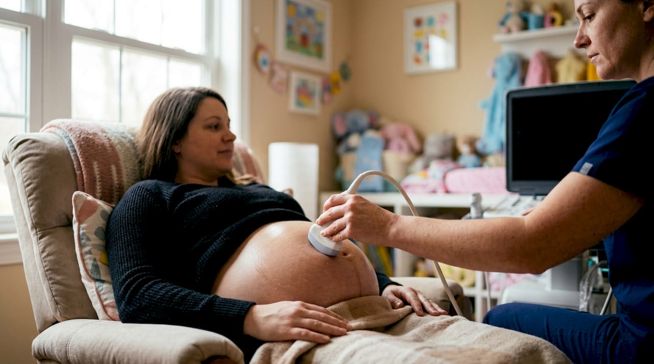
4D ultrasound adds one more layer: real-time video. The same volume data is captured continuously, so parents can watch the baby yawn, blink, or move its hands. The distinction matters because 4D surpasses 3D when families want to see live movement rather than a single detailed still.
AI-enhanced portraits take the grayscale 3D image and convert it into a full-color, photorealistic rendering. The AI reads the actual fetal anatomy captured in the scan, so the portrait reflects your baby's real facial structure rather than a generic face blend. Parents can also customize skin tone, hair color, and ethnicity to better reflect family features. These are creative previews, not medical predictions. Genetics, not software, determines what your newborn will actually look like.
Several factors affect image quality before the AI even gets involved:
- Fetal position: A baby facing the placenta or spine blocks the face from the probe.
- Amniotic fluid: Clear fluid around the face acts like a natural window. Low fluid reduces detail.
- Placenta location: An anterior placenta sits between the probe and the baby, reducing signal clarity.
- Gestational age: Too early means not enough fat under the skin. Too late means less room to maneuver.
Pro Tip: Drink 64–80 ounces of water daily in the week before your scan. Better hydration improves amniotic fluid clarity, which directly improves image quality.
When is the best time to get a 3D fetal portrait?
Timing is the single biggest factor parents can control. The optimal window is 26–32 weeks, with 28 weeks as the sweet spot for most pregnancies. At that point, the baby has developed enough subcutaneous fat to give the face a full, rounded appearance, but the womb still has enough amniotic fluid and space for the baby to move into a favorable position.
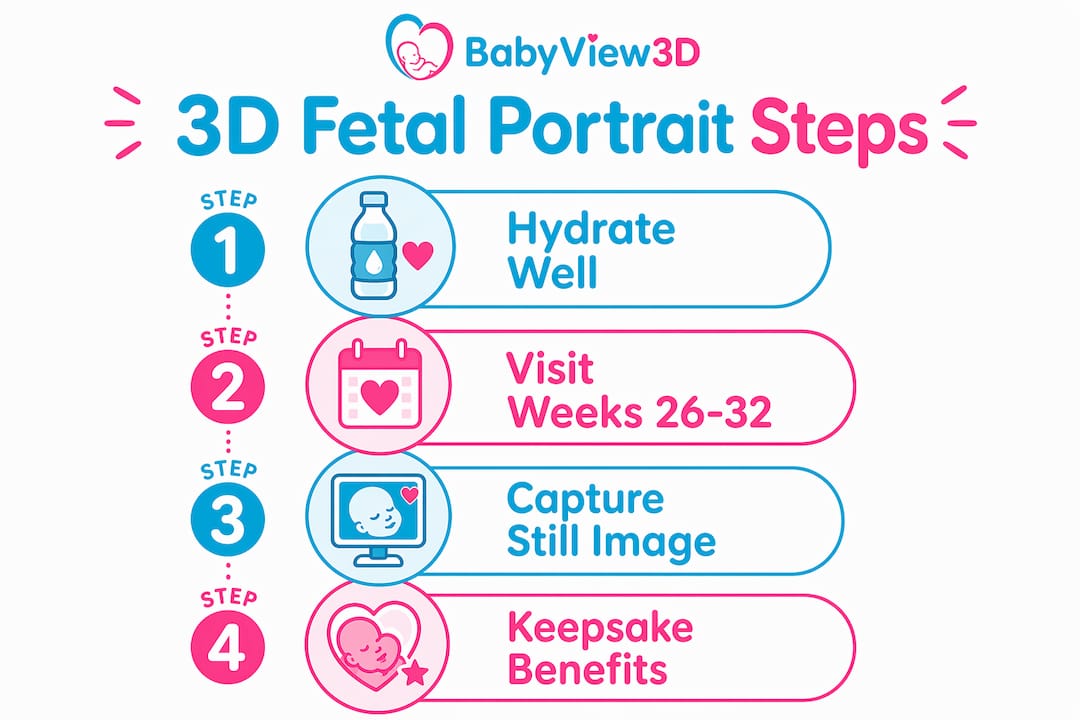
Before 26 weeks, the baby's face looks thin and skeletal because the fat layer has not filled in yet. After 32 weeks, the baby drops lower into the pelvis and the reduced space makes it harder to capture a clear facial view. Both scenarios produce images that are technically accurate but visually less satisfying for parents.
Here is how to time your session for the best results:
- Schedule between 26 and 32 weeks. This is the window recommended by most certified sonographers for elective 3D baby scans.
- Aim for 28 weeks if possible. The balance of fat development, fluid volume, and fetal mobility peaks around this point.
- Book earlier if you have an anterior placenta. Sonographers often recommend scheduling at 24–28 weeks in these cases, before the baby grows too large to reposition around the placenta.
- Avoid scheduling right after a meal. A full stomach can make the baby less active, reducing the chance of a clear facial view.
- Give yourself a reschedule option. Even expert sonographers face challenges when fetal positioning is unfavorable. Booking at a studio that offers a complimentary rescan removes the pressure.
Pro Tip: Drink a small glass of cold water or juice 15–20 minutes before your appointment. The temperature change often encourages the baby to shift position, improving the chance of a clear face shot.
How do 3D fetal portraits differ from 2D and 4D ultrasounds?
Parents often arrive at their first elective session unsure of what they have actually booked. The three scan types serve different purposes, and understanding the differences sets realistic expectations.
| Type | Image format | Primary use | Timing | Medical or elective |
|---|---|---|---|---|
| 2D ultrasound | Flat, grayscale cross-section | Anatomy screening, fetal heartbeat | Throughout pregnancy | Medical |
| 3D fetal portrait | Static, surface-rendered still image | Keepsake, bonding, AI portrait base | 26–32 weeks | Elective |
| 4D ultrasound | Real-time 3D video | Watching fetal movement | 26–32 weeks | Elective |
A 2D scan is the diagnostic standard. Ob-gyns and maternal-fetal medicine specialists use it to measure fetal growth, check organ development, and screen for structural anomalies. The image looks like a grainy cross-section because it is: a single flat slice through the body.
A 3D fetal portrait is a surface rendering. It shows the outside of the baby, not the inside. That makes it excellent for seeing facial features but not useful for diagnosing internal anatomy. 3D and 4D scans are elective keepsakes, not substitutes for the anatomy scan your ob-gyn orders.
4D is essentially 3D in motion. The same surface rendering updates in real time, producing a video rather than a still image. Families who want to see their baby move, smile, or suck a thumb typically prefer 4D sessions. Families who want a single detailed portrait to print, frame, or use as the base for an AI rendering typically prefer 3D.
What are the benefits of 3D fetal portraits for expecting parents?
The primary benefit is emotional. Seeing a lifelike image of your baby's face before birth creates a concrete, personal connection that a flat grayscale scan rarely achieves. Research consistently links 3D imaging to stronger prenatal bonding between parents and their unborn child.
The benefits extend beyond the parents themselves:
- Family sharing: Grandparents, siblings, and extended family who cannot attend the appointment can receive a digital image that feels personal and real, not clinical.
- Keepsake value: High-resolution 3D portraits can be printed, framed, or included in baby books. AI-enhanced color versions make particularly striking keepsakes.
- AI portrait customization: AI adjusts skin tone, ethnicity, and hair color to reflect family features, making the portrait feel more personal and emotionally resonant.
- Preparation: Some parents report that seeing detailed facial features helps them feel more prepared and less anxious about the birth.
- Accessibility: Digital files can be shared instantly with family members across the country or internationally.
Cost is a real consideration. Standard 3D/4D ultrasound sessions typically run $99–$200 depending on the studio and package. AI-enhanced portrait rendering is usually an add-on, priced at $9–$40 per render depending on the vendor. That puts a complete experience, scan plus AI portrait, within reach for most families without requiring a significant financial commitment.
One boundary matters: a 3D fetal portrait is a keepsake, not a diagnostic tool. It does not replace the anatomy scan your ob-gyn orders at 18–20 weeks. Treat it as a complement to your prenatal care, not a substitute.
How to get a high-quality 3D fetal portrait
Getting a great portrait requires more than just showing up. The session outcome depends on preparation, studio choice, and what you do with the files afterward.
- Choose a certified studio. Look for studios staffed by registered diagnostic medical sonographers (RDMS). Certification signals training and accountability.
- Understand the elective nature. Elective studios are not medical facilities. They do not diagnose conditions. Their job is to capture the best possible image of your baby's face.
- Budget for the full experience. Plan for $99–$200 for the scan session and an additional $9–$40 if you want an AI-enhanced color portrait.
- Hydrate consistently in the days before. Hydration improves amniotic fluid clarity, which directly affects how sharp the portrait looks.
- Request your original digital scan file. Original digital files preserve depth data that photos of printouts lose. If you plan to use an AI portrait service, the digital file produces a far more accurate result.
- Ask about rescan policies. Fetal positioning is unpredictable. Studios that offer a free return visit if the baby does not cooperate give you a real safety net.
Key Takeaways
A 3D fetal portrait is a still, surface-rendered image of your unborn baby's face, best captured between 26 and 32 weeks, and it serves as an emotional keepsake rather than a medical diagnostic tool.
| Point | Details |
|---|---|
| Definition | A 3D fetal portrait is a static, surface-rendered image built from multiple 2D ultrasound slices. |
| Best timing | Schedule between 26 and 32 weeks, with 28 weeks as the ideal gestational age. |
| Not a medical scan | 3D and 4D portraits are elective keepsakes and do not replace your ob-gyn's anatomy scan. |
| AI enhancement | AI converts grayscale 3D images into full-color portraits in 30–90 seconds using actual fetal anatomy. |
| File quality matters | Always request the original digital scan file for the most accurate AI portrait rendering. |
What I have learned after years of watching families experience 3D scans
The technology has changed faster than most parents realize. When 3D ultrasound first became widely available to expecting families, the images were grainy, shadowy, and required a lot of imagination to appreciate. Today, HD Live rendering and AI portrait tools produce images that genuinely look like photographs of a newborn. That shift has changed the emotional weight of the experience entirely.
What has not changed is the unpredictability. I have seen families arrive at the perfect gestational week, fully hydrated, with a cooperative baby, and still walk away with a blurry image because the cord was draped across the face. I have also seen families at 30 weeks with an anterior placenta get stunning portraits because the baby turned at exactly the right moment. You can control preparation. You cannot control the baby.
The AI portrait trend is the development I find most interesting right now. Ultrasound-anchored AI portraits are structurally constrained to the actual fetal anatomy from the scan, unlike generic face-blending apps that guess based on parent photos. That distinction matters. The portrait you get from a real scan file reflects your baby's actual facial structure, not a statistical average. It is a meaningfully different product.
My honest advice: book at 28 weeks, hydrate well, choose a studio with a rescan policy, and keep your expectations grounded. The goal is a meaningful memory, not a perfect photograph. When the conditions are right, these portraits are genuinely extraordinary. When they are not, a rescan a week later often solves the problem.
— LENIER
Bbview3d: see your baby before birth
Bbview3d has spent more than 15 years helping expecting families across the United States see their babies in stunning detail before birth. The studio offers elective 3D, 4D, and 8K HD Live ultrasound sessions staffed by certified sonographers, with packages designed to fit different budgets and gestational stages.
Families can browse session packages and pricing directly on the Bbview3d website, where first-appointment discounts are currently available. AI-enhanced portrait add-ons are included in select packages, giving parents a full-color, photorealistic image built from their actual scan data. The Bbview3d gallery shows real examples of what these portraits look like, so you know exactly what to expect before you book.
FAQ
What is a 3D fetal portrait exactly?
A 3D fetal portrait is a static, surface-rendered image of an unborn baby's face and features, reconstructed from multiple 2D ultrasound slices. It shows the baby's nose, lips, and cheeks in three-dimensional detail rather than a flat cross-section.
How accurate are 3D fetal portraits?
Image accuracy depends on fetal position, amniotic fluid clarity, placenta location, and gestational age. AI-enhanced portraits built from original digital scan files are more accurate than those generated from photos of printouts, because the digital file preserves the full depth data.
What is the difference between 3D and 4D ultrasound?
A 3D ultrasound produces a single still image of the baby's surface features. A 4D ultrasound captures the same surface rendering in real time, creating a video that shows fetal movement.
How much does a 3D fetal portrait cost?
Standard 3D/4D ultrasound sessions typically cost $99–$200. AI-enhanced color portrait add-ons range from $9–$40 per render depending on the studio and service provider.
Can a 3D fetal portrait replace my anatomy scan?
No. A 3D fetal portrait is an elective keepsake and does not replace the diagnostic 2D anatomy scan ordered by your ob-gyn at 18–20 weeks. The two serve completely different purposes.