Prenatal ultrasounds are one of the most powerful tools you have during pregnancy, yet most parents walk into their first appointment with little idea of what scan they are getting or why. Understanding the types of prenatal ultrasounds available, formally categorized under the broader field of obstetric sonography, gives you the ability to ask better questions, follow your provider's recommendations with confidence, and fully appreciate what each image reveals about your baby. This article breaks down every major scan type, the technology behind each one, and how to decide what is right for your situation.
Table of Contents
- Key takeaways
- 1. How the types of prenatal ultrasounds actually differ from each other
- 2. Early pregnancy scans: dating and viability checks
- 3. Mid-pregnancy scans: the anatomy and fetal anomaly scan
- 4. Doppler ultrasound: measuring blood flow
- 5. 3D and 4D ultrasound: advanced visualization
- 6. Emerging technology: wearable ultrasound patches
- 7. Third-trimester growth scans and their limits
- 8. Choosing the right scan: a side-by-side comparison
- My honest take on prenatal ultrasound choices
- See your baby in detail with Bbview3d
- FAQ
Key takeaways
| Point | Details |
|---|---|
| Probe type matters early on | Transvaginal probes give clearer images in the first trimester when abdominal views fall short. |
| Dating accuracy changes by trimester | Crown-rump length at 6 to 14 weeks dates a pregnancy within 3 to 8 days, far more precisely than third-trimester measurements. |
| Anomaly scans have real limits | Detection rates vary by condition, so understanding follow-up options prepares you for what happens next. |
| 3D and 4D scans serve dual purposes | Advanced imaging supports both medical evaluation and the emotional experience of seeing your baby in detail. |
| New technology is expanding options | Wearable ultrasound patches are now being studied for continuous fetal monitoring in high-risk pregnancies. |
1. How the types of prenatal ultrasounds actually differ from each other
Before comparing specific scans, you need one core concept: not all prenatal scans differ just by timing. They differ by probe type, imaging dimension, frequency setting, and clinical goal. Grouping them only by week of pregnancy misses the point.
The two probe categories you will hear most often are transabdominal and transvaginal. A transabdominal probe glides over your abdomen with gel. A transvaginal probe is inserted vaginally and sits much closer to the uterus, which means higher resolution images early in pregnancy. Importantly, probe choice depends on image quality and clinical need rather than gestational age alone.
Beyond probe type, scans also differ by what they measure:
- Standard 2D ultrasound produces flat, grayscale images and is the workhorse of routine prenatal care
- Doppler ultrasound adds blood flow data to standard imaging
- 3D ultrasound reconstructs still, three-dimensional surface images
- 4D ultrasound shows 3D images in real time, essentially a live video
- Specialized scans such as fetal echocardiography focus on specific organs
Timing also shapes what each scan can accomplish. The first trimester is optimal for dating accuracy. The second trimester is the window for anatomy screening. The third trimester is used to check growth, position, and placental function.
Pro Tip: Ask your provider before every appointment what type of scan they are performing and what specific clinical question it is designed to answer. This one habit will make every prenatal visit more meaningful.
2. Early pregnancy scans: dating and viability checks
The first scan most parents receive falls between 6 and 14 weeks. Its primary job is to confirm the pregnancy is viable, establish a due date, and check for multiple pregnancies. This is where crown-rump length measurement becomes the gold standard for gestational dating, accurate within 3 to 8 days when performed between 8 weeks and 13 weeks 6 days.
If your provider cannot get a clear abdominal view, they may switch to a transvaginal probe. Higher-frequency transvaginal probes deliver superior resolution in early pregnancy and improve structural detection rates compared to transabdominal imaging alone. This is completely routine and not a cause for concern.
What you can expect at an early scan:
- Confirmation of heartbeat and number of embryos
- Measurement of the gestational sac and crown-rump length
- Early check of the uterus and ovaries
- Nuchal translucency measurement if done at 11 to 14 weeks as part of first-trimester screening
The nuchal translucency scan is a specific type of early prenatal screening test that measures fluid at the back of the baby's neck. Combined with blood work, it screens for chromosomal conditions like Down syndrome. This is a separate purpose from a simple dating scan, even though both happen in the same trimester.
Pro Tip: Drink water before a transabdominal early scan to fill your bladder. This lifts the uterus forward and improves image clarity. You may be told to empty your bladder if a transvaginal approach is needed instead.
For parents preparing for their first ultrasound appointment, knowing what type of scan is planned ahead of time reduces anxiety significantly.
3. Mid-pregnancy scans: the anatomy and fetal anomaly scan
The anatomy scan, typically performed between 18 and 22 weeks, is the most detailed routine ultrasound in pregnancy. Routine prenatal care includes this fetal anomaly scan as a standard offering, designed to examine dozens of fetal structures in a single appointment.
Here is what a sonographer reviews during this scan:
- Brain and spine, including screening for spina bifida
- Heart chambers and major vessels
- Kidneys, stomach, and bladder
- Facial features including lip formation
- Limbs, hands, and feet
- Placental location and amniotic fluid levels
- Umbilical cord structure
This is also the scan where sex can be confirmed if you want to know.
Here is the part many parents are not told upfront. Detection rates vary significantly by condition. Spina bifida is detected approximately 98% of the time, but major heart defects are identified in only around 58% of cases at this scan. Some conditions simply do not present visibly at 18 to 20 weeks, which is why false negatives in anomaly screening are a known limitation, not a failure of the sonographer.
Pro Tip: If the sonographer finds something unclear, ask specifically what a follow-up scan would look for and when it should happen. Vague findings are common and often resolve, but you deserve a clear protocol, not just reassurance.
| Imaging type | Best use at this stage | Key limitation |
|---|---|---|
| Standard 2D anomaly scan | Full structural survey of fetal anatomy | Some defects not yet visible at 20 weeks |
| Doppler flow study | Placental blood flow, growth concerns | Requires trained interpretation |
| 3D ultrasound | Detailed surface imaging of face and limbs | Not a replacement for 2D diagnostic scans |
4. Doppler ultrasound: measuring blood flow
Doppler ultrasound is a distinct category within different fetal imaging technologies. It does not just show anatomy. It measures the speed and direction of blood moving through vessels, which standard 2D imaging cannot do.
Your provider may order Doppler imaging if there are concerns about fetal growth restriction, placenta function, or high-risk conditions like preeclampsia. The umbilical artery Doppler is the most common application. It tells your provider whether blood is flowing normally from placenta to baby. Uterine artery Doppler can identify abnormal placental circulation earlier in pregnancy.
Cerebral Doppler, which checks blood flow in the fetal brain, is used when growth restriction is already confirmed. This combination of types of placental imaging scans and cerebral assessments gives providers a detailed picture of how your baby is compensating under stress.
5. 3D and 4D ultrasound: advanced visualization
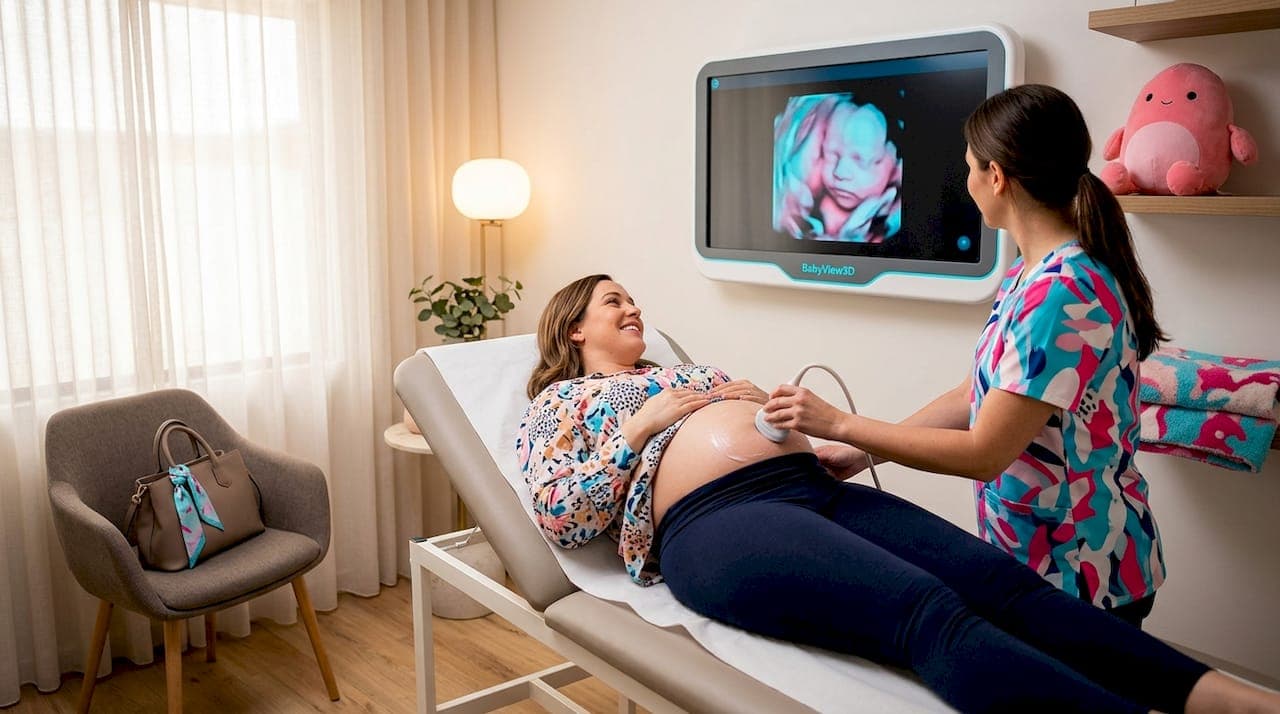
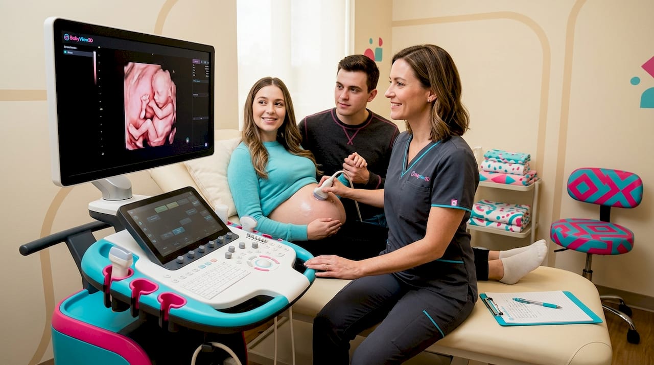
3D and 4D scans represent a significant leap in what parents can see and what providers can evaluate. A 3D scan reconstructs surface images from multiple 2D slices, giving a lifelike view of your baby's face, hands, and body contour. A 4D scan adds the time dimension, showing those images as moving real-time video.
Medically, 3D imaging assists in evaluating cleft lip, skeletal anomalies, and some cardiac conditions where surface structure matters. For parents, the experience of seeing a baby yawn, stretch, or bring their hand to their face creates a bond that flat 2D imaging simply cannot replicate. You can learn more about the 3D ultrasound process to understand what a typical session involves.
The optimal window for 3D and 4D imaging is generally between 26 and 32 weeks. Before 26 weeks, there is not enough subcutaneous fat for clear surface rendering. After 32 weeks, the baby is often too large and positioned in ways that obscure the face.
6. Emerging technology: wearable ultrasound patches
One of the most significant developments in real-time fetal imaging tools is the wearable ultrasound patch. This is not science fiction. A multi-center study published in 2026 showed that wearable ultrasound patches can monitor fetal heart rate and other parameters continuously over hours, with measurement agreement close to standard clinical devices.
The clinical implication is substantial. Standard prenatal ultrasounds are snapshots taken at single appointments. Conditions like growth restriction or placental insufficiency can develop and worsen between visits. Continuous fetal monitoring via wearable patches could help detect complications that a monthly scan would miss entirely.
These patches are currently most applicable in high-risk pregnancies and hospital settings, but fetal imaging software advances are moving quickly. Understanding this technology today helps you ask informed questions as it becomes more accessible.
7. Third-trimester growth scans and their limits
Growth scans in the third trimester serve a specific purpose: tracking whether your baby is growing at the expected rate for their gestational age. They also check amniotic fluid volume, fetal position, and placental grading.
What many parents do not realize is that dating accuracy drops considerably late in pregnancy. Second-trimester dating carries a margin of error of 7 to 10 days, but third-trimester measurements can be off by 21 to 30 days. More importantly, third-trimester biometric measurements should not be used alone to revise your due date. A baby measuring small late in pregnancy may be experiencing growth restriction, not a dating error. Your provider needs to distinguish between those two possibilities before changing any clinical plan.
8. Choosing the right scan: a side-by-side comparison
Different scans serve different purposes, and knowing which to prioritize depends on where you are in pregnancy and what questions your provider is trying to answer.
| Scan type | Typical timing | Primary purpose | Key consideration |
|---|---|---|---|
| Dating scan (transabdominal or transvaginal) | 6 to 14 weeks | Confirm viability, establish due date | Crown-rump length most accurate early |
| Nuchal translucency | 11 to 14 weeks | Screen for chromosomal conditions | Combined with blood test for best accuracy |
| Fetal anomaly scan | 18 to 22 weeks | Full structural survey | Some defects not detectable at this stage |
| Doppler study | Varies, often 24+ weeks | Blood flow in umbilical or cerebral vessels | Used for growth or placental concerns |
| 3D or 4D scan | 26 to 32 weeks optimal | Surface imaging, parental bonding | Not a diagnostic replacement for 2D |
| Growth scan | 28 to 36 weeks | Monitor size, fluid, and position | Do not use to revise due dates late in pregnancy |
| Wearable ultrasound patch | High-risk pregnancy use | Continuous monitoring between appointments | Emerging technology, not yet universally available |
When you meet with your provider, ask these questions to guide your decisions:
- What specific clinical question is this scan addressing?
- What will happen if something unclear is found?
- Is 3D or 4D imaging available and appropriate for this appointment?
- Are there any high-risk factors in my pregnancy that call for additional types of scans?
My honest take on prenatal ultrasound choices
I've spent years watching expecting parents walk into ultrasound appointments without knowing whether they are getting a dating scan, an anomaly scan, or a Doppler study. They leave reassured but unaware of what was actually checked, and more importantly, what was not.
My experience tells me the biggest missed opportunity in prenatal care is the gap between what a scan is designed to do and what parents assume it does. Many parents believe the 20-week anomaly scan is a full clearance. It is not. A major cardiac defect has only around a 58% detection rate at that visit. That is not a criticism of the technology. It is the reality of fetal development timelines.
What I've observed consistently is that parents who understand the purpose of each scan ask better follow-up questions and feel less blindsided if something unexpected comes up. Knowing that a transvaginal probe is simply a better tool early in pregnancy, not a sign of a problem, removes unnecessary worry.
The technology advances I find most interesting are not the 3D renderings, impressive as they are. The wearable continuous monitoring patches represent a real shift in how we think about fetal surveillance. Right now prenatal care is built around scheduled snapshots. Continuous monitoring could close the gaps that matter most in high-risk situations.
Talk to your provider at every appointment. Ask what they are looking for. Ask what would prompt a follow-up. You are not being difficult. You are being a good advocate for your baby.
— LENIER
See your baby in detail with Bbview3d
At Bbview3d, the focus is on giving your family an experience that goes beyond a standard clinical scan. With over 15 years of experience and certified sonographers, Bbview3d offers 3D, 4D, and HD Live imaging that lets you see your baby with stunning clarity before they arrive. Whether you are looking to complement your medical care with advanced visualization or create a keepsake memory your family will treasure, the prenatal ultrasound services at Bbview3d cover a range of options designed around your experience. First-time visitors can take advantage of a special introductory offer. Browse the services page and see what is available near you.
FAQ
What are the main types of prenatal ultrasounds?
The main types include the dating scan, nuchal translucency scan, fetal anomaly scan, Doppler ultrasound, 3D or 4D scans, and growth scans. Each serves a different clinical purpose depending on trimester and pregnancy risk factors.
When is a transvaginal ultrasound used instead of transabdominal?
A transvaginal probe is used when the abdominal view does not provide sufficient image quality, most often in early pregnancy. The decision is based on image clarity and clinical need, not gestational age alone.
How accurate is the dating scan?
Crown-rump length measurement between 8 and 14 weeks dates a pregnancy within 3 to 8 days, making it the most accurate gestational dating method available. Accuracy decreases significantly in the third trimester.
Can the 20-week anomaly scan detect all birth defects?
No. Detection rates vary by condition. Spina bifida is detected approximately 98% of the time, while major heart defects are found in roughly 58% of cases at this scan. Some conditions develop after the standard screening window.
What is the best time for a 3D or 4D ultrasound?
The clearest images typically come between 26 and 32 weeks, when the baby has enough fat under the skin for surface rendering but is not yet too large or positioned in a way that blocks the face.