
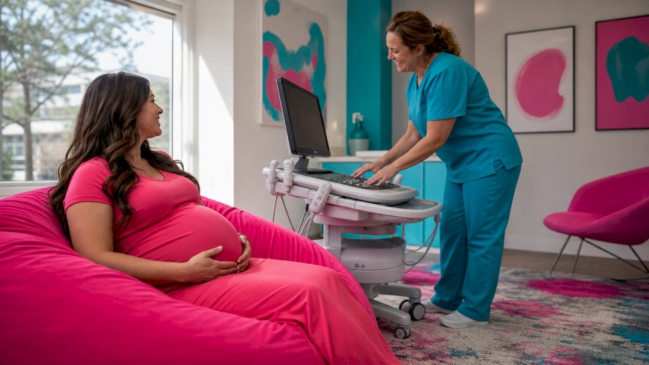
Ultrasound baby appearance varies because 3D scans are computed surface renders, not photographs. The imaging software reconstructs your baby's face from reflected sound waves, then applies algorithmic processing to produce the image you see on screen. Fetal position, gestational age, amniotic fluid levels, and placenta placement all shape the final result. Understanding why babies look different in ultrasound helps you interpret those images realistically and reduces the anxiety that comes with seeing an unexpected or unfamiliar face on the monitor.
Why do babies look different in ultrasound images?
The core reason is technical. 3D ultrasound images are computed surface renders built from thousands of reflected sound wave data points, not optical snapshots. The rendering algorithm fills gaps, smooths surfaces, and applies a color wash to create a recognizable face. When something blocks the signal, like a hand, the umbilical cord, or the placenta, the software either skips that data or misinterprets it. That misinterpretation is why some babies appear to have stretched features, flattened noses, or what parents often describe as an "alien" look.
Color is another source of confusion. The pink or yellow tones you see in a 3D scan are artificial rendering effects, not your baby's actual skin color. The software applies a visual color wash to make the image more appealing and easier to read. No ultrasound technology currently captures true in-utero skin tone. Knowing this prevents unnecessary worry about what the image suggests about your baby's complexion or features.
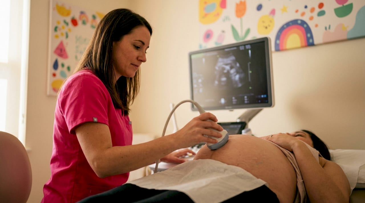
Pro Tip: Ask your sonographer to explain what you are seeing in real time. A brief verbal walkthrough of the image turns a confusing scan into a meaningful moment.
How fetal position shapes what you see
Fetal position is the single biggest variable in ultrasound image quality. A baby facing directly toward the probe produces the clearest facial images. A baby facing away, tucked chin-down, or pressed against the uterine wall produces a partial or distorted view.
Common position problems include:
- Face turned away from the probe. The back of the skull appears instead of facial features.
- Hands covering the face. Rendering algorithms can misread hands near the face as part of facial structure, creating distorted shapes.
- Chin pressed to chest. The nose and forehead compress, making features look flattened or merged.
- Umbilical cord crossing the face. The cord creates a shadow or artifact that interrupts facial detail.
- Baby positioned deep in the pelvis. Distance from the probe reduces signal strength and image sharpness.
Sonographers use several techniques to improve position. They may ask you to walk, drink cold water, or lie on your side to encourage the baby to shift. A repositioning attempt adds a few minutes to the session but can dramatically change the result. Experienced operators recognize position problems quickly and adjust the probe angle or ask for a short break before retrying.
Pro Tip: Drink 16–20 oz of water about an hour before your appointment. Hydration increases amniotic fluid volume, which gives the rendering software more acoustic space to work with.
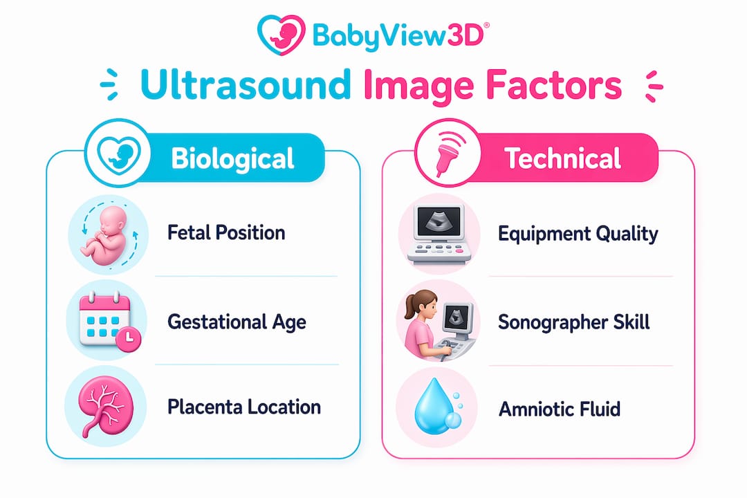
What gestational age does to image clarity
Timing is the factor most parents underestimate. Optimal 3D facial imaging occurs between 26 and 32 weeks gestation because the baby has enough subcutaneous fat to define facial features without being too crowded to photograph well.
Here is what changes at each stage:
- Before 26 weeks. The face lacks fat deposits, so the skin sits close to the bone. Images look skeletal rather than soft, and features appear sharp and angular in ways that do not reflect the newborn's actual look.
- 26–32 weeks. Fat fills in the cheeks, lips, and nose bridge. The baby has room to move and fluid surrounds the face. This window produces the clearest, most recognizable images.
- 33–36 weeks. The baby grows larger and the uterus becomes crowded. Good angles become harder to find, and the face is often pressed against the placenta or uterine wall.
- After 36 weeks. The baby's head typically engages in the pelvis. The probe cannot reach the face easily, and fluid levels drop, reducing image quality further.
The 26–32 week window is not just a scheduling preference. It is the period when fat volume, fluid space, and fetal size align to produce images that most closely resemble what the newborn will look like at birth. Booking outside this window is the most common reason parents are disappointed with their scan results.
For a deeper look at timing for facial detail, the gestational window matters more than any other single factor.
Technical and environmental factors that cause image differences
Beyond position and timing, the physical environment inside the womb directly affects image quality. Amniotic fluid acts as an acoustic window: the software needs at least 1–2 cm of clear fluid between the probe and the baby's face to render a sharp image. Without that fluid pocket, the algorithm cannot distinguish baby skin from the placenta or uterine wall, and the result looks blurry or artifact-heavy.
Placenta placement is a major structural variable. Anterior placenta placement sits on the front wall of the uterus, directly between the probe and the baby. This obstruction reduces signal clarity and limits useful imaging angles, particularly after 32 weeks. Research shows placenta site accounts for 18% of image quality variability in 3D scans. That is a substantial share of the outcome sitting entirely outside the parent's or sonographer's control.
| Factor | Effect on image | Controllable? |
|---|---|---|
| Amniotic fluid level | Low fluid causes blurring and artifacts | Partially (hydration helps) |
| Anterior placenta | Blocks probe access to baby's face | No |
| Fetal position | Determines which features are visible | Partially (repositioning techniques) |
| Gestational age | Affects fat volume and crowding | Yes (schedule in the 26–32 week window) |
| Equipment quality | Determines resolution and rendering accuracy | Yes (choose a facility with modern equipment) |
| Sonographer skill | Affects ability to minimize all other factors | Yes (choose an experienced operator) |
Sonographer skill and equipment quality substantially affect the final image. An experienced operator reads the acoustic environment quickly, adjusts probe pressure and angle, and knows when to pause and retry. Facilities using HD Live technology or 8K resolution systems produce noticeably sharper renders than entry-level equipment. The factors behind image quality are worth understanding before you book a session.
Pro Tip: Ask any facility what equipment generation they use before booking. HD Live and 8K systems produce significantly more detail than standard 3D setups from five or more years ago.
How to interpret ultrasound images and manage your expectations
A 3D ultrasound predicts facial geometry, not surface detail. Core features like nose bridge, lip shape, and cheek fullness often match the newborn's face closely. Fine details like skin texture, eye color, and hair do not appear in any ultrasound image.
Parents commonly misread these normal image characteristics as problems:
- Flattened nose. Usually caused by the face pressing against the uterine wall or placenta, not the baby's actual nose shape.
- Wide or asymmetric eyes. The rendering algorithm applies equal lighting across the surface, which can make eye sockets appear larger or uneven.
- Blurry or incomplete features. Almost always a fluid or position issue, not a structural concern.
- Unusual skin tone. The color is artificial. Pink, yellow, and orange tones are rendering choices, not biological information.
- Distorted or elongated features. Typically caused by the umbilical cord or a hand near the face that the algorithm misread as facial tissue.
4D ultrasound adds a time dimension, showing movement in real time. Watching the baby yawn, blink, or suck a thumb adds context that a still image cannot provide. Even so, 4D imaging carries the same technical limitations as 3D. Motion can actually reduce image sharpness if the baby moves during a critical rendering pass.
3D souvenir scans are captures meant to create a connection, not diagnostic tools. Research shows 51% of 3D souvenir scans result in poor or failed image quality. That figure reflects how many variables affect the outcome, and it is a strong argument for choosing an experienced facility and booking at the right gestational age. Distorted images do not indicate anything wrong with the baby's health or development.
Key Takeaways
Ultrasound image variation is caused by technical and biological factors, not by differences in the baby's actual appearance, and understanding each factor helps parents interpret scans accurately.
| Point | Details |
|---|---|
| Computed renders, not photos | 3D scans are algorithmic reconstructions; color and texture are artificial effects. |
| Optimal timing is 26–32 weeks | Fat volume and fluid space peak in this window, producing the clearest facial images. |
| Placenta site matters | Anterior placenta accounts for 18% of image quality variance and cannot be changed. |
| Position drives clarity | A face-forward baby produces the best images; repositioning techniques can help. |
| Skill and equipment count | Experienced sonographers and modern HD Live or 8K systems reduce most controllable variables. |
What I've learned watching parents react to their first 3D scan
I have watched hundreds of parents go quiet when they see their baby's face for the first time on a 3D monitor. Most of the time, the silence is not wonder. It is confusion. The image looks nothing like what they expected, and they do not know what to say.
The most common reaction I hear is some version of "Is that normal?" The answer is almost always yes, and the explanation is simple once you understand that the image is a software reconstruction, not a window into the womb. But parents rarely arrive knowing that. They expect a photograph and get an algorithm.
What I find genuinely frustrating is how rarely facilities explain this before the scan starts. A 60-second briefing about fetal position, fluid levels, and the 26–32 week window would eliminate most of the anxiety I see in those first moments. Parents who understand what they are looking at enjoy the experience far more than those who are left to interpret a strange image on their own.
My honest recommendation is to book between 28 and 30 weeks, hydrate well beforehand, and choose a facility where the sonographer talks you through the image in real time. The scan itself is not the memory. The moment you recognize your baby's face is. Give yourself the best conditions to have that moment clearly.
— LENIER
See your baby clearly with Bbview3d
Bbview3d has spent more than 15 years helping families see their babies before birth with the clarity that makes the experience genuinely memorable. Certified sonographers guide every session, and the technology includes HD Live and 8K resolution systems that produce the sharpest renders available today.

If you are planning a session, the 3D and 4D ultrasound services at Bbview3d are designed around the factors that matter most: the right equipment, the right timing, and a sonographer who explains what you are seeing. Browse the image gallery to see what a well-timed, expertly captured scan actually looks like. First-appointment discounts are currently available at select locations.
FAQ
Why does my baby look weird in a 3D ultrasound?
Most unusual appearances come from fetal position, low amniotic fluid, or the umbilical cord near the face. The rendering algorithm misreads obstructions as facial tissue, producing distorted shapes that do not reflect the baby's actual features.
What is the best week for a clear 3D ultrasound?
The 26–32 week window produces the clearest images because the baby has enough fat to define facial features and enough fluid space for the software to render them accurately. Most specialists recommend booking between 28 and 30 weeks for the best results.
Does the baby actually look like the ultrasound image?
Core facial geometry, including nose bridge, lip shape, and cheek fullness, often matches the newborn's face closely. Skin texture, eye color, and fine surface details do not appear in any ultrasound image.
Does an anterior placenta affect ultrasound image quality?
Yes. An anterior placenta sits between the probe and the baby's face, blocking signal angles and reducing clarity. Placenta site accounts for 18% of image quality variance in 3D scans and cannot be repositioned.
Are the colors in a 3D ultrasound real?
No. The pink, yellow, or orange tones in a 3D scan are artificial color washes applied by the rendering software. They are visual effects designed to make the image easier to read, not accurate representations of the baby's skin color.