

Color imaging in ultrasound is defined as a technique that overlays color-coded blood flow data onto standard grayscale anatomical images, giving clinicians a real-time view of how blood moves through fetal vessels. Clinicians call this technique color Doppler ultrasound, and it is the recognized industry standard for prenatal cardiovascular assessment. The role of color imaging in ultrasound goes far beyond adding visual interest. It reveals blood flow direction, velocity, and patterns that a black-and-white scan simply cannot show. For expectant parents, understanding what those colors mean transforms a confusing image into a meaningful window into your baby's health.
How does color Doppler ultrasound work in prenatal imaging?
Color Doppler ultrasound works by detecting the frequency shift of sound waves bouncing off moving red blood cells. When blood moves toward the probe, the returning sound waves arrive at a higher frequency. When blood moves away, the frequency drops. The scanner translates these frequency shifts into color, producing the vivid overlay you see on screen.
The color coding follows a standard called BART: Blue Away, Red Towards. Blood flowing toward the probe appears red. Blood flowing away appears blue. This is the single most important thing to understand about color ultrasound images. The colors do not represent oxygenated versus deoxygenated blood, and they do not reflect the actual color of blood inside the body.

Color Doppler always works on top of a standard B-mode grayscale image. The grayscale layer shows anatomy, such as the heart chambers, vessels, and surrounding tissue. The color layer adds functional information about what is happening inside those structures. Together, they give the sonographer both a map and a live traffic report.
Key technical factors that shape what you see on screen include:
- Probe angle: The beam must strike blood flow at an angle to detect movement. A probe held at 90 degrees to a vessel will show no color at all, even in a healthy vessel.
- Gain settings: Too much gain creates color noise outside the vessels. Too little gain misses slow-moving flow.
- Scale and PRF (pulse repetition frequency): These settings control the velocity range the machine can detect. Setting them too low causes a color artifact called aliasing.
- Vessel depth: Deeper vessels produce weaker signals, which can reduce color clarity.
Pro Tip: If you see the colors on screen change frequently during your scan, that is normal. The sonographer is actively adjusting settings to get the clearest possible picture of your baby's blood flow.
What benefits does color imaging provide over traditional ultrasound?
Color Doppler ultrasound provides benefits that standard grayscale scans cannot replicate. Real-time blood flow visualization significantly aids fetal cardiovascular health assessments, giving clinicians a functional view alongside the structural one. A grayscale scan can show that the heart has four chambers. Color Doppler can show whether blood is actually moving through those chambers correctly.
The clinical benefits for expectant parents include:
- Fetal heart screening: Color Doppler identifies abnormal flow patterns in the fetal heart early, flagging potential structural issues for follow-up.
- Umbilical cord assessment: Sonographers use color flow imaging to confirm blood is moving properly through the umbilical arteries and vein, which directly reflects placental function.
- Brain and organ perfusion: Color imaging shows blood reaching the fetal brain and major organs, supporting assessments of fetal growth and wellbeing.
- Placental location: Color Doppler helps identify placenta previa or abnormal placental attachment by mapping blood vessel distribution.
Beyond the clinical picture, color imaging carries real emotional weight. Watching blood pulse through your baby's heart in real time is a different experience from viewing a static gray outline. Many parents describe it as the moment the pregnancy feels undeniably real. Prenatal imaging has well-documented emotional benefits for families, and color Doppler adds a layer of visible, living proof that your baby's cardiovascular system is working.
Color Doppler also serves as a qualitative screening tool before more detailed spectral Doppler analysis. When a sonographer spots a color pattern that looks unusual, they can immediately zoom in with spectral Doppler to measure exact velocities. This two-step approach makes the entire examination more efficient and more thorough.
What are the key considerations and limitations of color imaging?
Color Doppler is powerful, but it has real limitations that every expectant parent should understand. Knowing these limitations prevents unnecessary worry when the image looks different from what you expected.
-
Angle dependency is absolute. Doppler accuracy requires the beam angle to stay below 60 degrees relative to blood flow. At 90 degrees, the machine detects zero velocity and shows no color, even in a perfectly healthy vessel. This is why your sonographer moves the probe constantly.
-
Aliasing is normal, not alarming. Aliasing occurs when blood velocity exceeds the machine's current PRF setting, causing colors to "wrap around" and appear reversed. It looks dramatic on screen but is a technical artifact, not a sign of a problem. The sonographer corrects it by adjusting the PRF or baseline.
-
Slow flow is hard to detect. Very small vessels and very slow-moving blood can fall below the machine's detection threshold. This is a known limitation of color Doppler and is why sonographers sometimes switch to power Doppler, which is more sensitive to low-velocity flow.
-
Gain errors create false impressions. Too much gain makes color bleed outside vessel walls, which can look alarming but means nothing clinically. Too little gain makes vessels appear empty. Sonographer skill in managing these settings is the single biggest factor in image quality.
-
Body habitus affects penetration. Tissue depth, maternal body composition, and fetal position all affect how well sound waves reach their target. Some scans will simply have less color clarity than others, and that reflects physics, not a health concern.
Pro Tip: Ask your sonographer to explain what you are seeing in real time. Most are happy to walk you through the color patterns, and understanding the image reduces anxiety significantly.
How does color imaging complement 3D and 4D ultrasound?
Color Doppler and volumetric ultrasound technologies serve different purposes, and they work best together. Understanding the difference helps you get more out of every prenatal appointment.
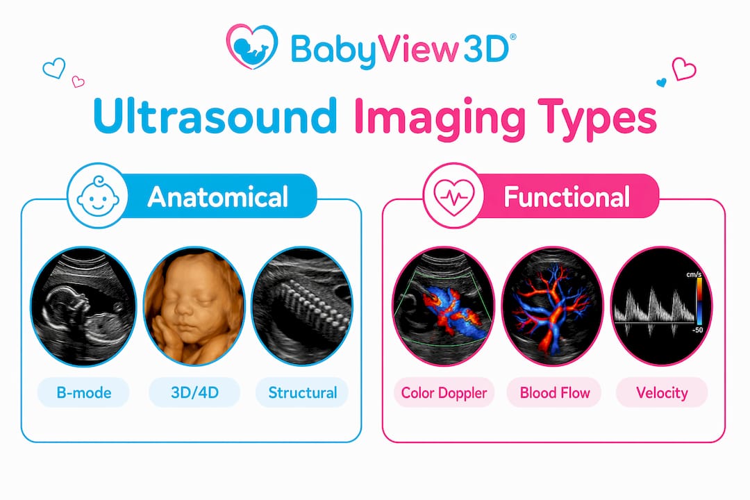
3D and 4D ultrasounds capture volumetric data to produce detailed structural images of the fetal face, limbs, and body. 3D produces a static volume. 4D adds real-time motion, so you can watch your baby yawn, stretch, or open their eyes. These modalities answer the question: what does my baby look like?
Color Doppler answers a different question: how is my baby's blood moving? It adds functional information on top of the anatomical picture that 3D and 4D provide.
| Imaging type | Primary purpose | What parents see |
|---|---|---|
| B-mode (2D grayscale) | Anatomical structure | Organs, measurements, fetal position |
| Color Doppler | Blood flow direction and velocity | Color-coded vessels and heart chambers |
| 3D ultrasound | Static volumetric structure | Detailed facial and body features |
| 4D ultrasound | Real-time volumetric motion | Baby moving, facial expressions |
A complete prenatal assessment often uses all of these tools in the same session. The sonographer starts with B-mode to orient the anatomy, adds color Doppler to assess cardiovascular function, and may use 3D or 4D to give parents a detailed view of their baby's features. Each modality adds a layer of information the others cannot provide. Understanding the types of prenatal ultrasounds available helps parents ask better questions and feel more confident in what they are seeing.
The combination is especially valuable for fetal heart screening. A 4D view shows the heart's structure and motion. Color Doppler confirms that blood is flowing through each chamber and valve in the right direction. Together, they give clinicians far more confidence than either modality alone.
Key Takeaways
Color Doppler ultrasound is the standard tool for assessing fetal blood flow, and understanding how it works helps expectant parents interpret their scans with confidence rather than confusion.
| Point | Details |
|---|---|
| Colors show flow direction, not blood type | Red means blood flows toward the probe; blue means it flows away. Neither color reflects oxygenation. |
| Angle and settings drive image quality | Sonographers adjust probe angle, gain, and PRF constantly to produce accurate color images. |
| Aliasing is a normal artifact | Color wrap-around on screen is a technical setting issue, not a sign of a health problem. |
| Color Doppler complements 3D and 4D imaging | Volumetric scans show structure; color Doppler adds functional blood flow data for a fuller assessment. |
| Sonographer skill is the key variable | The quality of color Doppler results depends heavily on the experience and technique of the person performing the scan. |
What I have learned watching color imaging change prenatal care
I have spent years watching expectant parents react to color Doppler images, and the pattern is consistent. Parents walk in expecting a black-and-white picture. They walk out having watched their baby's heart pump blood in real time. That shift matters more than most clinical discussions acknowledge.
The most common misunderstanding I see is the red-equals-oxygenated assumption. Parents see red on the screen and assume it marks healthy, oxygen-rich blood. When the color shifts to blue, they worry. Taking 30 seconds to explain the BART principle eliminates that anxiety entirely. Sonographers who explain as they scan produce calmer, more engaged parents.
Color Doppler also changed how I think about diagnostic confidence. A grayscale image of a fetal heart is a snapshot. Color Doppler turns it into a live performance. Watching flow patterns in real time gives clinicians information that no static image can replicate. The color flow imaging purpose is not decoration. It is a functional diagnostic layer that makes every prenatal scan more informative.
My honest advice to every expectant parent: do not watch the screen in silence. Ask questions. Ask what the colors mean. Ask why the sonographer is moving the probe. The more you understand what you are seeing, the more meaningful the experience becomes. A prenatal ultrasound is one of the few moments you get to observe your baby before birth. Color imaging makes that observation richer, and you deserve to understand every part of it.
— LENIER
Bbview3d brings color Doppler technology to your prenatal experience
Bbview3d has delivered premium prenatal ultrasound experiences for over 15 years, with certified sonographers who use advanced imaging technology at centers across the United States. Color Doppler is part of how Bbview3d's team assesses fetal cardiovascular health while giving families a vivid, real-time view of their baby.
Every session combines clinical accuracy with the emotional experience families remember. Bbview3d's packages include HD Live imaging, 3D and 4D volumetric scans, and visual keepsakes that capture your baby's earliest moments. Certified sonographers guide you through every image so nothing on screen feels confusing or worrying. Visit Bbview3d's prenatal services to see available packages and book your appointment, including a first-visit discount for new families.
FAQ
What does color Doppler ultrasound show during a prenatal scan?
Color Doppler shows the direction and velocity of blood flow through fetal vessels and the heart, overlaid on a standard grayscale anatomical image. It does not show tissue structure or fetal features the way 3D or 4D imaging does.
Why do the colors on my ultrasound keep changing?
The sonographer is continuously adjusting probe angle, gain, and scale settings to optimize the image. Color changes during a scan are a normal part of the process, not a sign that something is wrong.
Do the red and blue colors mean oxygenated and deoxygenated blood?
No. Red indicates blood flowing toward the probe, and blue indicates blood flowing away from it. The colors reflect flow direction relative to the transducer, not blood oxygen content.
Is color Doppler ultrasound safe during pregnancy?
Color Doppler is non-invasive and widely used for fetal and maternal health monitoring. It uses the same sound wave technology as standard ultrasound, with no radiation involved.
How is color Doppler different from a 4D ultrasound?
Color Doppler maps blood flow dynamics inside vessels and the heart. 4D ultrasound captures real-time volumetric motion of the fetal body and face. The two techniques provide different types of information and are often used together in the same prenatal session.